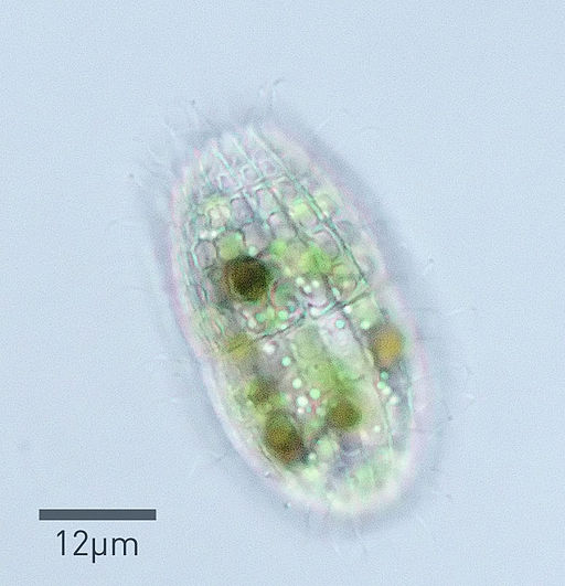
What is the Phylum Ciliophora?
Overview
Also known as ciliated protozoans, the phylum Ciliophora consists of single-celled organisms within the subkingdom Protozoa that move by means of cilia. Currently, there are over 7,500 described species within the group. However, it is suspected that there are many others yet to be identified.
Some of the species are free-living organisms found in different habitats around the world while others exist as parasites of vertebrates, invertebrates, and some mammals.
Some examples of the phylum Ciliophora include:
- Spirostomum ambiguum
- Strombidium lagenula
- Paramecium caudatum
- Paramecium aurelia
- Spirostomum minus
Classification of the Phylum Ciliophora
Domain: Eukaryota - Characterized by membrane-bound organelles
Kingdom: Protista - Single-celled protozoa (Ciliophora is sometimes placed in the kingdom Chromista which comprises unicellular and multicellular eukaryotic species)
Subkingdom: Protozoa - Single-celled heterotrophs of the kingdom Protista
Phylum: Ciliophora - Ciliated protozoans
Subphyla:
- Spirostomum minus
- Spirostomum minus
Characteristics
Morphology
As mentioned, phylum Ciliophora species (ciliates) are single-celled organisms. They greatly vary in size and shape depending on the species. For instance, while members of the class Colpodea range between 10 and 500 um in length, others, like Geleiids can be as large as 4 mm in length while some Stentor species are about 2 mm in length. As such, they can be seen with the naked eye.
Apart from size, different species also exhibit significant variations in the general morphology/shape. Some of the species have an ovoid to ellipsoid shape with rounded posterior and anterior ends. Sonderia species are good examples of ciliates with this morphology.
Some of the other distinct shapes include; kidney-shaped ciliates (e.g. Colpodea species), loop-shaped ciliates (e.g. Geleiids), funnel and worm-shaped species (e.g. Spirostomum species), and pear-shaped species with one pointed end (e.g. some Caenomorphia and Äcineto species). These differences in shape and size have made it easier to identify the type of ciliate.
Nuclei
Like many other protozoa, phylum Ciliophora species do not have a cell wall. However, they have a flexible cell membrane that consists of amphipathic lipids. The membrane encloses a number of organelles that include two nuclei. In addition to the cell membrane, ciliates also have a flexible pellicle. It mainly consists of protein and other components and is located below the cell membrane.
Here, it has been shown to help maintain the shape of the ciliate. Because they have two nucleus types, Ciliophora species are described as having nuclear dimorphism.
Here, the two nuclei are macronucleus (polyploid macronucleus) and micronucleus (diploid micronucleus). The macronucleus consists of multiple sets of chromosomes and is therefore referred to as being highly polyploidy (has more than two sets of homologous chromosomes).
As the name suggests, it's also larger in size and generally contains many copies of genes of the genome. Also known as the somatic nucleus, the polyploid macronucleus is involved in the vegetative growth of the cell.
Compared to the micronucleus, the macronucleus is transcriptionally very active with higher rates of gene transcription. This helps in the maintenance of the large cytoplasm found in phylum Ciliophora and is suggested to be the basis of the large size of ciliates. Compared to the diploid macronucleus, the micronucleus is smaller in size and not very transcriptionally active.
As a diploid nucleus, it contains a double set of chromosomes which explains the smaller size. While it is not very transcriptionally active, the micronucleus, also known as the gametic nucleus, becomes active during sexual conjugation. As such, it allows individual cells to exchange genetic material when mating.
* Essentially, the macronucleus regulates RNA synthesis and consequently cellular regulation.
* While the macronucleus is larger than the micronucleus, it's formed through the fusion/karyogamy of micronuclei daughter cells.
* Macronucleus usually divide through amitosis while micronucleus divide through meiosis.
Cilia
One of the defining characteristics of all phylum Ciliophora species is the fact that they all have hair-like structures known as cilia. Here, however, it's worth noting that cilia may be absent at certain stages of the organism’s life cycle.
In different types of species, these structures are arranged in rows of kinities (in lines). Whereas cilia are dense in some of the species, the distribution is sparse in others.
For instance, Cronotheridium and some Stentor species are characterized by a dense distribution of cilia on the cell body. The distribution is more sparse among some Acineta and Calipera species.
Among members of the genera Acineta and Cothurina among others, these hair-like structures are likely to be distributed at the anterior portion of the cell body with very little to no cilia at the posterior end.
In some of the species, cilia are arranged in a manner that forms a distinct organelle. For instance, compact cilia may form cirri or membranelles. Being a thicker structure than cilia, cirri are normally used for motility (by crawling) over surfaces. As such, it acts like a tiny limb-like structure that some caterpillars use to crawl over surfaces.
Membranelles are arranged around the mouth part (cytostome) of the organism where they aid in directing current and thus food material. Here, the series of membranelles forms the adoral zone of membranelles and help sweep food particles into the cytostome. Moreover, they have been shown to promote motility in some species.
* Cilia are generally used for motility.
Basal bodies
Like cilia, basal bodies are important microtubules found in phylum Ciliophora species. The number of basal bodies varies between different species in the group. For instance, whereas an average-sized Tetrahymena may have 900 basal bodies distributed at different parts of the cell body, a large Paramecium cell could have as many as 5,000 basal bodies.
In some species, some of these bodies are longer than others. However, this is largely dependent on where they are located on the Ciliophora. In a Paramecium, the longest basal bodies (about 600 nm in length) are located around the oral apparatus while the shorter ones (about 330 nm) can be found at the posterior part of the organism.
* Basal bodies are arranged along the longitudinal rows of cilia.
* The basal bodies are mainly located along the cortical rows as well as the oral apparatus.
* Basal bodies are made up of 9+3 microtubule blades.
The basal bodies serve to nucleate and anchor cortical and oral apparatus cilia. As such, they play a vital role in the motility and feeding process of the organism.
Ingestion and Digestion of Food Material
Some of the parts involved in the ingestion and digestion of food material include the oral groove, buccal overture, the cytostome, and food vacuoles. Through the beating action of cilia, many phylum Ciliophora species are able to move water over the surface of the cell which allows them to draw in food particles.
Here, cilia lining the oral groove not only help in creating these water currents but also sieve through the food particles and move them to the buccal overture and into the cytostome (unwanted materials are expelled). In Ciliophora like Paramecium, the buccal overture is a small opening that leads to the buccal cavity.
Along with the vestibule, located between the oral groove and buccal cavity, the vestibule serves to pull in food material into the cytopharynx before reaching the cytostome which acts as the cell mouth.
From the cell mouth, these materials are moved to the food vacuole where they are acted upon by various digestive enzymes. Waste products are then removed through the anal canal known as the cytoproct.
In some cases, food materials are taken into the cell through phagocytosis. In Paramecium, for instance, the extension of the cell membrane encloses the food particle. These materials are then internalized and contained in the food vacuoles (phagosome) which in turn fuses with lysosome for digestion to take place.
Respiration - The exchange of gases occurs through the membrane and the semi-permeable pellicle. Here, gases move through the membrane through diffusion where carbon dioxide and waste like ammonia is released into the surrounding water, and oxygen obtained from the water taken into the cell.
Habitat
Different groups within the phylum Ciliophora have different lifestyles and can therefore be found in different habitats. While the majority of species can be found in aquatic environments, some are terrestrial and can be found in soil. Some species are parasitic while others form symbiotic relationships with their hosts.
Aquatic Habitats
The majority of free-living phylum Ciliophora species can be found in freshwater and marine habitats.
Freshwater Ciliophora (found in ponds, rivers, lakes, and springs, etc.) - Some of the species found include some members of the groups Oligotrichia, Stichotrichia, Peniculia, Karyorelictea, and Nassophorea among others. In freshwater environments, they occupy different niches and habitats that are conducive to their way of life.
For instance, Loxodes species are commonly found in benthic and planktonic niches where they feed on bacteria and smaller protists. Stentor and Blepharisma species, on the other hand, are planktonic.
Here, many species are active swimmers and feed randomly (as filter feeders of smaller protists and bacteria). However, some of the species remain attached to various substrates and feed on the materials around them.
Colpidium, Tetrahymena, Euplotes, and Apidisca species are largely benthic and thus commonly found deeper in the water where they exist as predators of bacteria and other protists.
* While many phylum Ciliophora species move by means of cilia, some are not highly mobile and remain attached to various substrates. These ciliates are known as stalked ciliates and exist as solitary organisms or in colonies. Many of these species resemble a tulip with a thin stalk attached to the substrate and broader upper portion.
Cilia are located at the upper portion of the body where they beat to create currents that draw in bacteria. In the event of poor conditions, many of the species can detach from the substrate and swim (with or without the stalk) to a more favorable location.
Marine ciliates - Euplotes, Tintinnids, and Uronema species are some examples of marine Ciliophora. Here, they can be found in the coastal niches to the ocean floor where they exist as free-living organisms and a few as parasites. Some of the species are planktonic and therefore swim or float around feeding on bacteria and some protists. These include the species, Cyclotrichium meunieri and Myionecta rubra.
As compared to planktonic species, benthic species are commonly found in association with other marine organisms (as commensals or as parasites). For instance, Haematophagus megapterae can be found in the baleen plates (comb-like strictures made up of keratin used to filter water during feeding) of humpback whales while Trichodina multidentis lives as an ectocommensal in the gut of some sea urchin. Trichodina species, on the other hand, are parasitic and cause disease and even death of various marine fish.
Terrestrial ciliates - Currently, there are an estimated 1,000 terrestrial ciliates. These species can be found in soil, especially in decomposing litter layers where they exist as decomposers. Here, however, they still feed on bacteria and other smaller protists in their surroundings.
Symbiotic ciliates - Many symbiotic Ciliophora species can be found in the gut of various animals. As mentioned, some of the species can be found in the gut of sea urchins in marine habitats. However, there are many others that can be found in the gut of herbivores.
Here, they benefit the host (herbivore) by breaking down various plant matter to release extra energy. In turn, they benefit from a conducive environment with a constant supply of nutrients. Aside from herbivores, they have also been identified in the alimentary tract of many other animals including hippos, gorillas, and chimpanzees, etc.
* Some of the most popular commensal in the rumen of ruminants are members of the Orders Entodiniomorphida and Vestibuliferida.
Parasitic Ciliophora - As opposed to commensals, parasitic Ciliophora benefit from their host and also cause harm and disease. In human beings, the species Balantidium coli is an example of a parasitic ciliate that causes disease.
Measuring between 50 and 200 um in length, the organism can invade the colonic mucosa of the gastrointestinal where it causes diarrhea and dysentery. Here, however, it's worth noting that human beings are accidental hosts of the ciliate (pigs are their primary hosts).
In aquatic habitats, Trichodinidae among other ciliates are parasites of fish.
Reproduction and Life Cycle
Phylum Ciliophora reproduces sexually and asexually. Whereas asexual reproduction occurs through binary fission, sexual reproduction involves conjugation between two individual cells. Conjugation/mating allows for the exchange and fusion of haploid micronuclei to produce a zygote nucleus. These cells then separate and divide to produce new daughter cells with genetic material from the two parents.
* The macronuclei are destroyed during the sexual mode of reproduction. However, they are again formed by micronuclei after new daughter cells are formed.
Some ciliates form cysts that allow them to survive harsh environmental conditions. For these cells, the cyst becomes part of their life cycle.
* Some of the species that form cysts are members of free-living Colpodida and parasitic Balantidium (B. coli).
Life Cycle of Balantidium coli
Cyst ingestion - Cysts are normally found in diarrhea stools of infected individuals. As such, they can be found in soil and contaminated water. They are smaller compared to the trophozoites (between 50 and 70 um in diameter) with a relatively thick wall and dark yellow in color. They are ingested when an individual consumes contaminated food or water.
Excystation - Following ingestion, excystation occurs in the small intestine. This process produces trophozoite forms that migrate to the large intestine. These forms are relatively larger, measuring 50 to 130 um in length and 20 to 70 um in width.
Compared to the spherical cysts, trophozoites have an ovoid morphology with a pointed anterior end. Like many other phylum Ciliophora species, they are covered in cilia that promote rotary motility. They are also characterized by a funnel-shaped mouthpart (systome) as well as contractive vacuoles that can be seen under the microscope.
Replication - In the large intestine and appendix, these trophozoites divide through binary fission. At the same time, they can undergo conjugation which allows for the exchange of genetic material.
Encystation - In the large intestine, some cells undergo encystation where a hard outer wall is formed. This prepares them for harsh environmental conditions when they are excreted along with fecal matter.
* In cases where trophozoite forms penetrate the colon wall and replicate, they can cause significant damage.
Significance of Phylum Ciliophora
As sources of food - In marine and freshwater habitats, Ciliophora feed on bacteria, some algae, and smaller protists. However, as part of the food chain, they are also a source of food for many other higher organisms in their surroundings.
As decomposers - Some of the free-living Ciliophora (especially terrestrial ciliates) are involved in the decomposition of various organic matter and thus contribute to the humus. This is particularly important in agriculture given that the process supplies nutrients needed for plant growth and development.
As symbionts - Various ciliophora species can be found in the gut of many animals where they help in breaking down various plant components. This process releases extra energy for the host.
Return from What is the Phylum Ciliophora? to MicroscopeMaster home
References
F. Verni & G. Rosati. (2011). Resting cysts: A survival strategy in Protozoa Ciliophora.
Gianna Pitsch et al. (2019). Seasonality of Planktonic Freshwater Ciliates: Are Analyses Based on V9 Regions of the 18S rRNA Gene Correlated With Morphospecies Counts?
Lynn D.H. (2016) Ciliophora. In: Archibald J. et al. (eds) Handbook of the Protists.
Wilhelm Foissner, Tony Charleston, Martin Kreutz, and Dennis Gordon. (2012). phylum Ciliophora. New Zealand inventory of biodiversity, p 217132.
Links
https://www.accessscience.com/content/ciliophora/136300
https://www.bionity.com/en/encyclopedia/Ciliate.html
Find out how to advertise on MicroscopeMaster!


