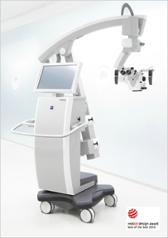
Surgical Microscope
Buyer's Guide
Zeiss, Leica & Others
- Important Features & Innovations -
Originally a radical concept, the surgical microscope has become an indispensible tool since the first recorded use of a lighted binocular microscope during an operation in 1922.
First used in neurosurgical procedures, this tool is now heavily utilized in the surgical practice of ophthalmology, otolaryngology, plastic and reconstructive medicine, dentistry, gynecology, and urology.
The simple purpose of this microscope is to improve the surgeon’s view and the usual components of microscopy, magnification, resolution, and illumination are important.
However, new aspects such as stability, size, viewing and recording capabilities, positioning, and the ability to integrate with image guided surgical tools must be addressed in designing and using this tool.
Important Features
The modern surgical microscope can be mounted on a stand, placed on a table top, or worn on the surgeon’s head. Some of them can be hung on the ceiling or wall to conserve floor space in the surgical suite.
- Binocular Head - A binocular head with adjustable eyepieces is basic, but the ability to add another head for an assistant, also known as a teaching head, allows two people to work with magnification in the operating field. The heads should independently pivot and have separate magnification levels. Ideally, this microscope head will have the flexibility to be retrofitted with a camera to document the procedure on an SD card or DVD as well as to allow colleagues to view the operation in real time.
- Foot Controls - for tilt, focus, and zoom are crucial because this will free both of the surgeon’s hands to continue the procedure without interruption.
- Illumination - is especially critical to maximize the capabilities of a surgical microscope. Incandescent, fiber optic, and halogen are the principle type of illumination used with these microscopes and the ability to accurately adjust the intensity is fundamental.
- Magnification - The extreme magnification found in laboratory microscopes is not necessary for a surgical microscope and is usually set in three to six presets ranging from 6X to 25X. Typically, a wide field of view is more helpful to a surgeon than extreme magnification.
Other factors to consider are antimicrobial-coated surfaces and internally routed cables. The latter is important because the controls, lighting, and documentation technology require wiring. Loose wiring gets in the way of the surgical team and can harbor bacteria.
Since this type of microscope is quite expensive, purchasing one that can be used by a variety of medical specialties is a cost effective solution.
Most of these instruments can be adapted to meet general surgical needs; however a surgical microscope used for ophthalmic procedures is designed differently.
On an ophthalmic microscope the binocular head is canted at an angle of as much as 45 degrees while the head on a microscope used for other procedures is straight.
Additionally, an ophthalmic surgical microscope has specialized lighting, focusing, and magnification requirements.
Zeiss Surgical Microscopes
The genesis of the modern surgical scope can be attributed to The Zeiss Corporation in 1953.
Early innovations and improvements focused on lighting methods, scalability of the microscope, and decreasing their size to limit intrusion into the surgical field.
Today the forefront of innovation is in the area of documentation and patient data storage, and fluorescence microscopy.
|
OPMI Pentero Zeiss has several multidisciplinary surgical microscope systems, but the OPMI Pentero series incorporates the newest available technology. This system can be ceiling mounted to be out of the way when it is not in use but touching an activation button will quickly lower it into position over the patient. It has instant magnification change, precise xy positioning, and can incorporate intraoperative fluorescence tools to assist the surgeon. |
Image of OPMI Pentero 900 from www.Zeiss.de |
The Infrared 800, Flow 800, and Blue 400 fluorescence tools immeasurably increase the usefulness of the Zeiss surgical microscope.
A fluorescence-based angiography tool, the Infrared 800 allows the surgeon to see the vascular circulation at the surgical site and determine the sequence and direction of blood flow with continuous color mapping.
The Flow 800 is the analytical tool used in conjunction with the Infrared 800. Using fluorescence video sequences generated by the Inflared 800, this tool allows the surgeon to compare before and after arterial function. This is very important in neurosurgical procedures.
The Blue 400 allows the surgeon to precisely target and resect tumors. This ability is critical in removing brain tumors because the surgeon certainly wants to completely excise the entire tumor but not unnecessarily remove healthy tissue. This is of special concern in the resection of brain tumors because all brain tissue is functional.
Using a special light source and appropriate filters, this fluorescence illumination tool accurately fixes and defines the edges of the tumor because the tumor fluoresces blue.
Another module is available for this microscope that exports the images from the microscope to a picture archiving and communications system for instant viewing at other terminals on the network. This information can be directly linked to the patient’s records.
Zeiss also makes the OPMI VISU series of microscopes for ophthalmic procedures as well as the OPMI Neuro and OPMI Vario system for minimally invasive spinal surgery.
Most of these microscopes can be integrated with image guided surgical tools and have different lighting options.
Touch screen controls are standard but foot controls may be added.
Leica Microsystems
As well as developing its own system of fluorescence microscopy, Leica designed the first head-mounted microscope.
- HM500 - designed for use in all types of surgeries. Besides the advantage of hands-free operation, this unit allows the surgeon the freedom to move around the patient. The surgeon can switch between a 9X magnification for higher resolution work to a 2X magnification for a wider view of the entire surgical field. Additionally, an integrated camera can document the procedure and display the surgeon’s view on a screen for the surgical staff assisting in the operation.
- Leica M520 and M720 systems - versatile and can be used in all surgical applications although ophthalmic procedures would be better served by using one of Leica’s microscopes specifically made for eye surgeries. These are the premium surgical microscopes heavily used in neurosurgical procedures. With motorized focus and magnification, these systems can be used with image guided surgical tools. Also,xenon arc illumination sources provide exceptionally bright illumination. One factor that is important for neurosurgical application is that the M720 has a redesigned optical head that is more compact than other heads.
- M844 ophthalmic microscope offers the standard features inherent in a high quality surgical microscopeas well asdirect halogen lighting for sharp contrast and accurate color at low intensity levels.
- Leica M320 series - a general microscope that targets the otolaryngology and dental markets. Even though this microscope has a small floor footprint, LED lighting, and easy maneuverability, this basic scope has less sophisticated controls than some models. However, it has internally routed cables and is easy to sterilize. Also, high-definition recording on an SD card is available for this unit.
- FL400 - Leica’s oncological fluorescence package that is adaptable to all its high-end surgical microscopes. The surgeon can quickly change from bright white light to blue fluorescence to see the outlines of the tumor. This system will also record the procedure in high definition.
- Leica Ultra Observer - with six observation ports, provides visualization for the surgeon, three assistants, and two video or camera adapters. Leica’s Dual Imagining technology allows display of patient data on the microscope and provides data correlation for image guided surgical techniques.
Other Manufacturers
Topcon
Topcon Medical manufactures two ophthalmic microscopes and a general purpose surgical scope. Some of their features include:
- LCD display Flare
- reducing optics
- Small footprint
- Programmable foot control
- Optional XY translator
- Teaching head
Visine Industries
Visine Industries is another company with a presence in the surgical microscope market.
These scopes have optional beam splitters and camera attachments, a selection of filters, and floor stand or table clamps.
Globe Surgical makes three dental microscopes and two otolaryngology microscope.
Basic microscopes lacking some of the sophisticated features of a Zeiss or a Leica, these units are small and can be hung from the wall or ceiling as well as mounted on a floor stand.
Nikon and Olympus
Two major microscope manufacturers, Nikon and Olympus, do not have a presence in this particular branch of microscopy.
Olympus does make several endoscopes to view the interior of body cavities and organs in a minimally invasive manner, but the company does not manufacture a true surgical microscope.
If leasing a microscope would be a more practical solution for you, Surgical Microscopes, LLC leases several models of Zeiss, Leica, and Topcon microscopes.
And Amazon...
The surgical microscope has graduated from being a luxury to an operating room necessity.
More than merely providing magnification, new microscopes can record and display patient data and remove the uncertainty from tumor resections by employing fluorescence microscopy.
Scalability is a key consideration in choosing a microscope to add flexibility and the ability to incorporate advances in this field of microscopy.
Take a look at Dental Microscopes and Endoscopes
Return from Surgical Microscope to Microscopy Applications
Return to Best Microscope Reviews and Research Home
Find out how to advertise on MicroscopeMaster!


