Cell Biology
Organelles, Cycles and Division, Signaling & Techniques
As a sub-discipline of biology, cell biology is concerned with the study of the structure and function of cells. As such, it can explain the structure of different types of cells, types of cell components, the metabolic processes of a cell, cell life cycle and signaling pathways to name a few.
Here, we shall look at some of the major areas of cell biology including some of the tools used.
Cell Theory
Cell Theory is a basic principle in biology that was formulated by Thodor Schwann, Matthias Schleiden and Rudolph Virchow.
According to the Cell Theory:
- All the living things (organisms) are made up of cells
- The cell is the basic unit of life
- Living cells come from existing/living cells
Recently, the theory was modified to include the following ideas:
- Energy flow takes place within cells
- Heredity information passes from one cell to another
- All cells have the same basic chemical composition
Cell Biology - The Cell
A cell is a basic unit of life. This simply means that a cell is the smallest unit of a living thing. While some organisms are only made up of a single cell (bacteria, yeast etc) others are multicellular organisms made up of many cells.
While there is a clear difference between unicellular and multicellular organisms, some organisms may transition from unicellular organisms to multicellular organisms under certain conditions.
A good example of this is slime mold that tends to transition to a multicellular organism under stressful conditions. However, they are simply described as being partially multicellular. Therefore, the cell is the basic building block of any given organism.
For a multicellular organism, cells are specialized, which means that they have differentiated to carry out given functions.
The following are examples of specialized cells:
Sperm Cells - Sperm cells serve to fertilize the female egg to form the embryo.
Red Blood Cells - Red cells contain a protein molecule known as hemoglobin and serve to transport oxygen to all parts of the body and expel carbon dioxide from the body.
White Blood Cells - There are different types of white blood cells that serve to protect the body from disease causing organisms.
- Basophils, Lymphocytes, Neutrophils, Monocytes, Eosinophils
Cardiomyocytes - These are cardiac muscle cells that make up the heart muscle.
Nerve Cells (neurons) - These are cells of the nervous system that transmit information to and from different parts of the body (information is transmitted as electric and chemical signals). See also Sensory Cells.
Cell Organelles
Any given cell will have three major components.
These include:
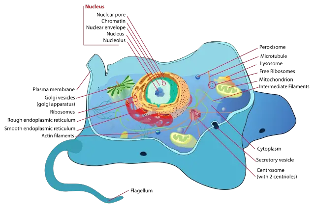
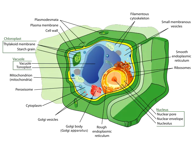
The cell wall is a complex, highly organized structure that defines the shape of a plant cell (it is also found in bacteria, fungi, algae, and archaea).
In addition to defining the shape of plant cells, a cell wall has a few other functions that include maintaining the structural integrity of a cell, acting as a line of defense against a variety of external factors as well as hosting various channels, pores and receptors that regulate various functions of a cell. As such, it is a multifunctional structure in plant cells that also contributes to plant growth.
Also known as the plasma membrane, the cell membrane is a bi-lipid membrane layer (it is a double membranous structure) that is also composed of proteins and carbohydrates. This fluid like structure encircles the cell thereby containing the contents of a cell.
It's also selectively permeable, which means that it only allows certain materials (nutrients and minerals etc) to pass through to sustain the cell. The cell membrane also functions to protect the cell and ensure stability.
The nucleus may be described as the largest organelle of a cell. The nucleus is itself surrounded by a double membrane (nuclear envelope) and contains genetic information (genes) making it the control center of a cell. As such, it controls such activities cell metabolism and reproduction.
The cytoplasm is the fluid matrix (jelly-like) found inside the cell (outside the nucleus). Various types of organelles and minerals (salts) are suspended in this constantly streaming fluid. Apart from containing all the cell organelles, the cytoplasm also helps maintain the shape of a cell.
See differences between cytosol and cytoplasm.
Cell organelles may be described as cell subunits specialized to carry out given functions within the cell. There are different types of organelles in cells that carry out given functions.
The following are some of organelles that can be found in a cell (excluding the cell membrane, cytosol and nucleus which are mentioned above):
Mitochondria - The mitochondria are rod-shaped organelles and sites of ATP synthesis. The mitochondria is also surrounded by a double membrane (with the inner membrane being highly folded forming the cristae).
This organelle is commonly referred to as a power- generator given that it converts oxygen and nutrients in to a chemical energy known as ATP (adenosine triphosphate) which provides the energy required for various activities of the cell.
Apart from being a site for ATP synthesis, the mitochondrion is also involved in the self-destruction of a cell in a process known as apoptosis.
Ribosomes - Found in the cytoplasm and the surface of the rough endoplasmic reticulum, ribosomes are composed of RNA and proteins. They may be described as the "cell factories" given that they are responsible for the synthesis of protein molecules.
Lysosomes - These are sac-like structures that are surrounded by a membrane (a single membrane). Lysosomes contain digestive enzymes, which are responsible for breaking down proteins, lipids and nucleic acids. In addition, lysosomes are also involved in the removal of waste molecules as well as recycling of molecular subunits.
Golgi body - These are flattened structures in a cell responsible for temporary storage of protein in the cell.
Vacuoles - Vacuoles are also enclosed by a membrane and function to store such material as food, water, minerals and waste products among others.
Some of the other organelles include:
- Plastids (in plants)
- Cilia and flagella (in animal cells)
- A central vacuole (in plants)
- Vesicle
- Chloroplasts (plants)
Cell Biology - Cell Cycle and Division
Cell cycle refers to a sequence in actively dividing cells where the cells pass through several stages before ultimately dividing.
The stages of cell cycle include:
- Two gap phases (G1 and G2)
- The S phase (synthesis)
- The M phase
At GI, the metabolic changes take place preparing the cell for the division process. At a given point known as the restriction point, the cell is committed to cell division and moves to the next phase.
S - The S phase involves DNA synthesis. It is during this phase that the replication of genetic material starts with each of the chromosome having two chromatic sisters.
G2 - During this phase, there are metabolic changes that assemble the necessary cytoplasmic materials for the mitosis process and splitting of the mother cell.
M - The M phase is where nuclear division takes place and followed by the division of the cell.
For most animals, cells may divide by mitosis or meiosis. While the two processes result in the production of new cells, they are different and produce different daughter cells.
Mitosis
Mitosis is the type of cell division that occurs in all somatic cells. These are the types of cells that make up the body tissues (apart from gametes/sex cells). Therefore, the primary role of mitosis is growth and replacing worn out cells.
Essentially, mitosis results in diploid cells from one cell. Here, the chromosome is copied followed by the separation of the copies on different sides of the cell before the cell ultimately separates into two. In the end, each of the new cells has a copy of the chromosome.
Mitosis has 5 major phases, which include:
Interphase - Here, the DNA strand is replicated/copied to produce what is known as a bivalent chromosome (consisting of two chromatids or DNA strands that are replicas of each other). During the interphase stage, the new strand is attached to the original one at a point known as the centromere.
Prophase - This is the second stage of mitosis. Here, the bivalent chromosomes formed during interphase condense to form tight packages.
Metaphase - This is the third stage where each of the chromosome line up at the center of the cell. The nucleus membrane has already started dissolving with each of the mitotic spindles attaching themselves to each of the chromatids. Here, it appears as if the chromatids are being stretched towards either pole of the cell.
Anaphase - During anaphase, the fourth stage of mitosis, the chromatids that had attached to the spindles are separated (the chromatids are separated from their copies) and pulled to either side of the cell. This results in two groups of monovalent chromosomes.
Telophase - At the end of anaphase, another stage starts where nuclear membranes start to form around the two formed groups of chromosomes. The spindle fibers that attached to the chromatids get disassembled. Here, the chromosomes also condense.
Eventually, the cytoplasm divides/splits with a cell membrane forming on each of the two daughter cells. This process is known as cytokinesis. Each of the new cells has 46 monovalent chromosomes and has identical genetic information as the other.
In mitosis, it's important that the same genetic information is copied when forming new cells. This is because the chromosomes have all the information concerning the function of the cell.
Successful copying of information on to the new cells ensures that the new cell functions properly. In the event that there is a problem, then the new cell will be unable to perform its function as it should be. This would result in complications depending on the function of the cell.
Meiosis
Unlike mitosis, meiosis produces haploid cells.
Diploid - Two new daughter cells from the original cell with the same number of chromosomes.
Haploid - With meiosis (a reductive type of cell division) the resulting cells will have less number of chromosomes.
Stages
Meiosis is also different from mitosis in that there are two phases of cell division. These are meiosis I and meiosis II.
Prophase 1 - Here, the homologous chromosomes pair and exchange DNA form recombinant chromosomes. This stage ends with the spindle fibers starting to form to attach to the chromosomes.
Metaphase 1 - The bivalent chromosomes arranges double row having attached to the spindle fibers.
Anaphase 1 - The homologous chromosomes (in each bivalent) are separated and move to opposite poles of the cell.
Telophase 1 - With the separation of the chromosomes, a nuclear membrane starts to form around the two groups of the chromosomes. This is followed by cytokinesis where the cell splits to form two new cells. This is again followed by meiosis II. Meiosis II follows the same process as meiosis I. However, this halves the number of chromosomes.
* Meiosis is an important process that results in genetic diversity.
What are the differences between Meiosis and Mitosis?
Cell Biology - Cell Differentiation
All cells originate from a single cell (a single fertilized egg). In cell differentiation, cells become specialized as the body develops. Apart from the single original cell (the fertilized egg), stem cells are also unspecialized. However, under certain conditions, they can differentiate to become specialized cells that serve a specific function(s).
Although the differentiated somatic cells are different in that they perform different functions, they contain the same genome. However, the different types of cells only express some of these genes, which results in the differences morphological and physiological between them.
Cell Biology - Signal Transduction/Signaling
In cells, signal transductions involve the transmission of molecular signals. This is particularly from the exterior of the cell to its interior for appropriate cell response. Signals (biochemical changes) may either come from the environment the cell is in or from other cells that trigger changes.
Cells have receptors on the surface of the cell, which receives the signal prompting a response. For a response to take place, the signal has to be transmitted across the cell membrane.
Some of the common intracellular messengers include:
- cAMP,
- cGMP,
- nitric oxide,
- lipids
- Ca2+ ions
Cell signaling is very important given that it helps control and maintain the normal physiological processes in the body. Different signaling processes will result in varying responses including cell differentiation, proliferation of cells as well as metabolism among others.
Cell Biology Techniques
Cell biology is largely concerned with the study of the structure and functions of cells (morphological and physiological). For this reason, a number of techniques have to be employed.
Some of the main cell biology techniques include:
- Tissue culture/Cell Culture
- Microscopy Imaging
- Staining
Tissue Culture
Cells and tissues can be cultured using
complex media. With cells and tissues from more complex organisms, the culture
media has to be more complex so as to provide the same environment as the
environment from which the cell/tissue was obtained.
As for the tissue, the culturing process also allows for single cells to be obtained from the tissue in question for more studies.
The culture process requires the following:
- A solid media - Agar media
- Growth media - This contains nutrients such as amino acids, vitamins, salts, glucose and growth factors among others.
Cell culture is an important technique given that it allows for only a sample (cells or tissue) to be used to learn more about the cells without the need to use the organism as a whole. This also gives scientists a great opportunity to study the cells under varying conditions.
See Also: Cell Culture
Microscopy Imaging
Microscopes have been used since the 1670s to observe
cells. Today, microscopes have become indispensable tools in cell biology. There are many more microscopy techniques today that have allowed for better viewing of cells.
In recent years, the world of microscopy has experienced advancements in imaging technologies enabling increased amounts of information for microscopic analysis.
Some of the most common techniques used in cell biology include:
- Bright field microscopy
- Fluorescence microscopy
- Immunofluorescence
- Time lapse microscopy
Staining
Staining goes hand in hand with microscopy.
Although it may be regarded as an important part of microscopy, staining is
itself very useful in cell biology. It allows for increased contrast
which in turn allows for scientists to view different parts of a cell clearly.
Although staining is highly useful when it comes to viewing specimen under the microscope, it cannot be used when a scientist wants to observe living cells.
Conclusion
Cell biology is an important discipline that has allowed for viewing and studying of cells for decades now. It has become particularly important to differentiate and determine different types of cells, cell processes as well as understanding of various diseases and illnesses associated with cell malfunctioning.
With advancements in various cell biology techniques, it is becoming easier to learn more about cells and cell processes for effective intervention where necessary.
More on Cells:
Eukaryotes - Cell Structure and Differences
Prokaryotes - Cell Structure and Differences
Protists - Discovering the Kingdon Protista in Microscopy
- Paramecium - Classification, Structure, Function and Characteristics
- Vorticella - Characteristics, Structure, reproduction and Habitat
Diatoms - Classification and Characteristics
Fungi - Mold Under the Microscope , Aspergillus type
Algae - Reproduction, Identification and Classification
Protozoa - Anatomy, Classification, Life Cycle and Microscopy
Bacteria - Morphology, Types, Habitat, looking at anaerobes, Eubacteria
Archaea - Definition, Examples, Characteristics and Classification
What are the Functions of Lipids, Proteins and Liposaccharides on the Cell Membrane?
Learn about Passive Diffusion Vs Active Transport
Learn about Serotype and Antigens
And Multicellular Organisms - Development, Processes and Interactions
Related and Interesting articles:
Gram Staining - Purpose, Procedure and Preparation
Endospore Stain - Understanding Definitions, Techniques and Procedures
Capsule Stain - Definitions, Methods and Procedures
Check out Beginner Microscope Experiments.
And more advanced microscopy experiments such as Trichomes and Microscopy, Parasites under the Microscope, Bone Tissue under the Microscope, Tissue Culture
What are the differences between a Plant Cell and an Animal Cell?
What are the Differences between Microbiology and Biochemistry?
Return from Cell Biology to MicroscopeMaster Research Home
References
Hausman, Geoffrey M. Cooper, Robert E. (2000). "Signaling Molecules and Their Receptors".
Karl-Hermann Neumann and Jafargholi Imani, Ashwani Kumar (2009) Cell Division, Cell Growth, Cell Differentiation.
Lodish, Harvey (2013). Molecular Cell Biology.
Shai Shaham (2006) Methods in cell biology.
Links
http://www.di.uq.edu.au/sparqcbecellintro
Find out how to advertise on MicroscopeMaster!


