What is Near Field Scanning Optical Microscopy?
General Overview
Also known as scanning near-field optical microscopy (SNOM), near field scanning optical microscopy (NSOM) is a technique that overcomes the diffraction limit by combining the strengths of atomic force microscopy (AFM) and optical fluorescence microscopy. In doing so, near field scanning optical microscopy has made it possible to get a higher resolution (up to about 30nm) than that offered by ordinary optical microscopes.
Fundamentals
In order to understand how near field scanning optical microscopy breaks Abbe's diffraction limit, it's important to understand the limitation of a typical optical microscope. While light microscopy has been a valuable tool in many fields over the years, allowing users to observe objects that are too small to be seen by the naked eye, the limit in optical resolution (diffraction limit of light) makes it virtually impossible to distinguish two separate objects at a given point.
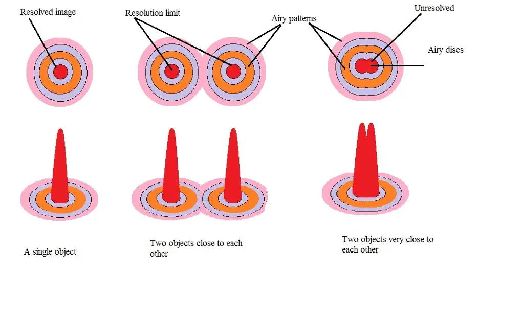
This means that even at the highest magnification possible, two separate points/objects would appear as one. Here, light originating from two objects that are close together scatter and overlap and thus appear as a single object.
The following is a diagrammatic image representing resolved and unresolved objects under a microscope:
Diagram (a)
In this image, it's possible to see that the microscope has no problem resolving a single object. While light from the object is scattered, it can be identified as a single object. As the objects get closer together, scattered light begins to overlap. However, it's still possible to identify that they are two different objects.
Eventually, when the objects get very close together and their scattered light completely overlaps, it becomes difficult to distinguish the two as they appear to be one object. While reducing the light wavelength and increasing the numerical aperture can help overcome this issue, it's worth noting that the wavelength of visible light falls between 400 and 700 nm. Therefore, regardless of how good the lens is, the microscope cannot resolve objects that are smaller than half the wavelength of light.
To overcome the limitations of an optical microscope, near field scanning optical microscopy uses an optical fiber probe which has an aperture that is smaller than the wavelength of light. This allows the microscope to offer a higher resolution (30nm in some cases).
This is largely dependent on the size of the aperture tip. Here, however, some of the aspects of near field scanning optical microscopy that makes this possible include an extremely (almost zero) working distance, longer scanning time, as well as lower intensity of the incident ray/light.
Probes and Aperture
As mentioned, near-field scanning optical microscopy uses fiber optic probes. These types of microscopes are known as aperture-type SNOM. The probes, which are aluminium coated, have a tapered geometry and thus have a pointed tip. This tip can be modified/tailored depending on application.
By coating the optical fiber with aluminium, this leaves a very small opening for light to pass through. During microscopy, this probe is placed very close to the sample so that the distance between the probe and the sample is only a few nanometers apart.
Currently, there are two main methods of preparing the optical fibers before they are coated with aluminium. One of the methods of preparing the fibers is known as heating and pulling. This method involves local heating by using carbon dioxide (CO2) gas laser. The fiber is then pulled apart.
The other method of preparing tapered optical fibers is known as chemical etching. Here, the fiber is dipped into a hydrofluoric acid covered with an organic solvent. In the process, the tip is formed at the meniscus which is itself formed at the interface between the solution and the solvent.
This method is generally preferred given that it makes it possible to reproduce probes at larger quantities through a single step. Moreover, it overcomes the problems associated with Turner's method including roughness of the glass surface.
Once the optical fiber is formed, the next step involves forming the aperture. In both chemical etching and heating and pulling, this is achieved by aluminium evaporation at an angle. As a result, deposition of aluminium at the apex/tip is smaller compared to the sides. Through this process, the probes produced have an aperture (ranging between 20 and 120nm in diameter) that is smaller than the wavelength of light.
Path of Light
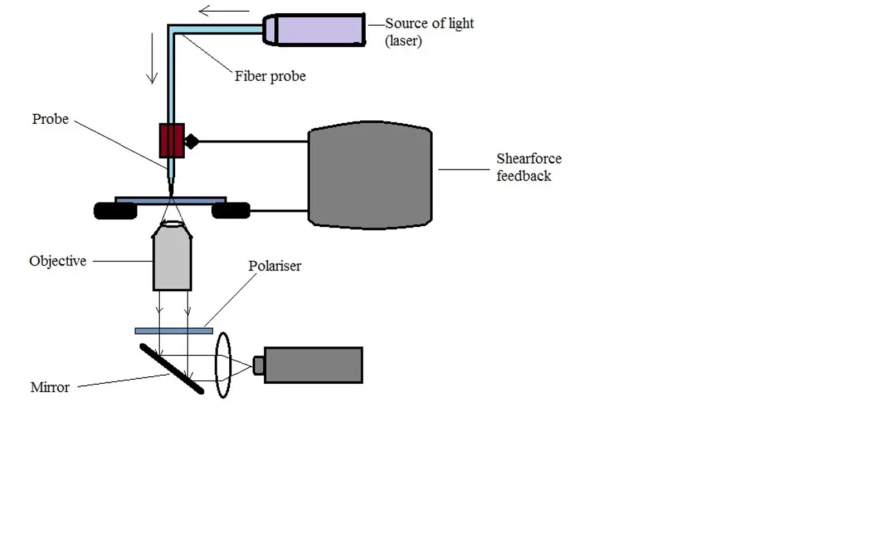
In a typical near-field scanning optical microscope, light (laser) is passed through the fiber optic and through the small aperture. To achieve sub-diffraction imaging, as mentioned, then the aperture of the probe has to be placed within the near-field region of the specimen in order to scan its surface. Given that far-field light is unable to pass through this small aperture, optical near-field is often used as it can penetrate through. As well, an evanescent field can also be used.
The size of the aperture plays a very important role in resolution as the intensity of light passing through is dependent on the size of the aperture. Therefore, by increasing or reducing the size of this aperture, resolution also changes with the change in light intensity.
Diagram (b)
Apertureless SNOM - Near-field imaging using near field scanning optical microscopy is also achieved using non-aperture SNOM tips.
This technique has been shown to produce better resolution when compared to Aperture-type SNOM (c-SNOM or collection SNOM). However, this method also has a disadvantage given that large far-field background signal from the light source results in signal detection. In this system, the sample-probe system is first illuminated globally so that the absorbed light (by the probe) is eventually emitted for near-field illumination of the sample.
In the event that the medium is excited below saturation, the emitted intensity of this probe has been inconstant. This is because as the light from the local sample is scattered, the intensity of excitation light is also modulated during the scanning process.
As the microscope tip (in c-SNOM) scans along the surface of the sample, the quality of the image obtained is governed by the diameter of the tip as well as its positioning rather than the diffraction limit associated with the typical microscope.
By coating the tip with aluminium, this helps minimize loss of light originating from the laser. This is particularly important given that this system allows for the light to be focused on specific points on the surface.
Generally, for this tip to be useful in Scanning Electrochemical Microscopy, studies have shown that the metal coating has to be electrochemically inert. This is one of the reasons metal coatings like gold and platinum are commonly used.
* The spatial resolution normally achieved in NSOM images is between 10 and 20 times better than the resolution obtained using ordinary light microscopes.
* Flat and smooth tips of the probe has been shown to help improve the quality of the image given that they significantly reduce the distance between the aperture and the sample - To flatten the tips, they are often polished using focused ion beam milling.
As already mentioned, a good resolution is achieved when the tip of the probe is very close to the sample surface. In order to keep the aperture near the sample, a type of force feedback known as shear force feedback is often used.
Here, the tip is moved above the sample surface in a horizontal manner where it experiences constant force. Generally, this involves oscillation of the probe using a piezoelectric device so that the tip moves from side to side.
With the tip approaching the sample, the shear force created lessens the intensity/frequency of these oscillations. This is detected and allows for the generation of a feedback signal which is in turn used to generate topographic images.
Application
Because of the high resolution achieved using near field scanning optical microscopy, the technique has a number of important applications including:
Fluorescence Imaging - A fluorescent microscope is a type of optical microscope that generally uses fluorescence to observe and study the characteristics of various substances.
Given that near field scanning optical microscopy has the capacity to image the distribution of labels down to the single fluorophore level, the technique has been employed in fluorescence imaging (fluorescence NSOM) thus producing high resolution optical imaging of various biological samples with fluorescent labels.
Being better suited for biological and biochemistry applications, this technique has been successfully used to investigate the properties of protein molecules including immunoglobin.
* In fluorescent NSOM, the molecules under investigations have to be labeled with appropriate dyes thus allowing them to be detected.
Near-field Raman Spectroscopy - In Near-field Raman Spectroscopy, near-field optics are combined with vibrational spectroscopies which results in molecular contrast n a lateral scale.
While this technique has been shown to have low efficiency, researchers have been able to achieve single-molecule sensitivity and sub-20 nm spatial resolution using the method. This technique is used for the purposes of studying the physical characteristics of biological specimen (e.g. proteins)
Check out a great page on Nanotechnology here
Scanning (SEM) - Learn about the SEMs high-resolution, three-dimensional images which provide topographical, morphological and compositional information making them invaluable in a variety of science and industry applications.
Transmission (TEM) - check out one of the most powerful microscopic tools available to-date, capable of producing high-resolution, detailed images 1 nanometer in size.
Cryo-Electron - is a type of transmission electron microscopy that allows for the specimen of interest to be viewed at cryogenic temperatures. Check it out.
Virtual - provides a simulated microscope experience via a computer program or Internet website for both educational and industrial applications and are easily operated and accessible.
Take a look at how Electron Microscopy compares to Super-Resolution Microscopy.
Taking a look at Viruses under the Microscope and answering the question, what are viruses?
As well as Atom under the Microscope and DNA under the Microscope
Electron microscopy (SEM and TEM) images of SARS-CoV-2 - Covid 19
Return to learning about the Nanonics Optometronic 4000SPM
Return from Near Field Scanning Optical Microscopy to MicroscopeMaster home
References
Bert Hecht, Beate Sick, Urs P. Wild, and Volker Deckert. (2000). Scanning near-field optical microscopy with aperture probes: Fundamentals and applications.
Heath A. Huckabay, Kevin P. Armendariz, William H. Newhart, Sarah M. Wildgen, and Robert C. Dunn. (2013). Near-Field Scanning Optical Microscopy for High-Resolution Membrane Studies.
Paul Bazylewski, Sabastine Ezugwu, and Giovanni Fanchini. (2017). A Review of Three-Dimensional Scanning Near-Field Optical Microscopy (3D-SNOM) and Its Applications in Nanoscale Light Management.
V. Sandoghdar. (2005). Near-Field Optical Microscopy.
Links
https://www.sciencemag.org/news/2011/04/scattering-microscope-peers-nanoscale
https://www.olympus-lifescience.com/en/microscope-resource/primer/anatomy/numaperture/
Find out how to advertise on MicroscopeMaster!


