Vorticella
Characteristics, Structure, Reproduction and Habitat
Overview: What is Vorticella?
Vorticella is a protozoa (protist) that belongs to the phylum Ciliophora. As such, they are eukaryotic ciliates that can be found in such habitats as fresh and salty water bodies among others.
According to studies, Vorticella is the largest genus of sessile peritrich ciliates with over 100 identified species. Depending on the habitat, this species will feed on a range of food material through the vestibule that acts as the entrance route for food.
Apart from being part of the food chain, it also plays a number of important roles in the environment that benefit human beings.
Some of the species in this genus include:
- V. campanula
- V. citrina
- V. marina
- V. communis
- V. striata
- V. utriculus
- V. sphaerica
Classification
Vorticella is a genus of ciliate protozoa that is classified under the following:
- Kingdom: Chromalveolata
- Phylum: Cilophora
- Superphylum: Alveolata
- Class: Oligohymenophorea
- Subclass: Peritrichia
- Order: Sessilida
- Family: Vorticellidae
Reproduction and Life Cycle
Asexual Reproduction
Like many other Infusoria (tiny aquatic organisms such as protozoa) division (binary fission) of the organisms into two (or sometimes more) daughter cells has been shown to be one of the means of propagation of Vorticella.
Before division of the organisms commences, the body of the organism shortens as it increases in breadth. This is then followed by the division of the nucleus where the macronucleus divides amitotically while the micronucleus divided mitotically. This is accompanied by the constriction of the organisms that ultimately divided the organism into two or more organisms.
While the parent organism divided to form two or more Vorticella, it is worth noting that only one of the new organisms retains the original stalk. This leaves the other to develop new cilia (temporary cilia) and ultimately a new stalk through which it can attach to another substrate or surface.
Binary fission of such Peritrichia as Vorticella has been shown to be unique compared to other ciliates given that it is often unequal and longitudinal (in that it runs along the oral-aboral axis of the organism).
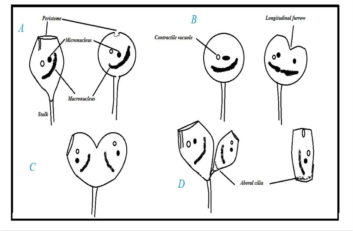
Diagrammatic representation of Longitudinal Binary Fission:
In this diagrammatic representation, it's possible to identify some of the parts of the organisms including: the peristome, micronucleus, macronucleus, a contractile vacuole, aboral cilia as well as the longitudinal furrow.
As binary fission begins, the peristome first closes up as the body (bell) of the organism shortens and increases in breadth (transverse elongation). The contractile pulsates during the division while the macronucleus shortens and condenses (as it moves in the middle of the cell transversely).
As division continues, constriction begins at the anterior end of the cell and gradually divided the cell down its length (from the peristome and towards the stalk). This ultimately divided the organism into two unequal parts (with one of the daughter cells being smaller).
Whereas the larger cell retains a stalk with which it can remain attached to a surface, the smaller one lacks a stalk but develops aboral cilia at its posterior. Compared to the larger cell with a stalk, the smaller cell becomes more cylindrical in shape and is known as a teletroch.
Using the aporal cilia, the cell then swims away and attaches to a surface through its scopula at its aporal end. The scopula is also responsible for producing a new stalk for the daughter cell that allows the organism to remain attached to the surface. Over time, the cylindrical shape develops into a bell shape as the organism matures.
* This division usually takes 20 to 30 minutes.
Sexual Reproduction/Conjugation
Apart from Binary fission, Vorticella have also been shown to reproduce through conjugation (sexual reproduction).
This is divided into two main phases that include:
Formation of micro and macro-conjugants
This phase involves a binary fission process that produces a larger and smaller cell (unequal cells). Here, the smaller cell (which may be more than one in some cases) is known as the micro-conjugant.
The micro-conjugants produced through this phase develop posterior cilia and having detached from the other larger cell, swim and attach to other surfaces. Compared to the telotrochs produced through asexual reproduction, micro-conjugants have been shown to be smaller in size. Moreover, micro-conjugants do not develop (metamorphose) in to adult forms and do not develop a stalk. After 24 hours, they die off rather than encyst as some of the telotrochs do during touch conditions.
Moreover, the larger cells that retained a stalk go through nuclear modifications and develop into macro-conjugants. In this state, the macro-conjugants can reproduce sexually with viable micro-conjugants.
Fusion
The second phase of sexual reproduction is commonly referred to as fusion of the conjugants. As the micro-conjugants swim about, they can come in contact with the macro-conjugants and attach to the lower part of the cell body near the stalk; Following this attachment, cilia of the micro-conjugants falls off followed by changes in the nuclear of the two conjugants.
Whereas the macronuclei of both conjugants degenerate (and absorb in the cell cytoplasm) the micronucleus in the macro conjugant undergo divisions that result in four micronuclei while those of the micro-conjugant divide several times to produce eight micronuclei.
In both conjugants, all the micronuclei apart from one degenerate so that each conjugant has a single micronucleus (female pronucleus of the macro conjugant and male pronucleus of the micronucleus).
The wall between the two conjugants then disappears allowing the two unite. This is then followed by fusion of the two pronuclei resulting in the formation of a zygote nucleus (synkaryon). Division of the zygote in turn produces seven macronuclei and a single micronucleus.
The micronucleus also undergoes another division to form two micronuclei that are separated following division of the cell. This results in one cell with one micronucleus and four macronuclei and the other with three macronuclei and one micronucleus.
The individual cells and micronucleus also continue with the division ultimately producing cells with one macro and micro nucleus.
The end result of these divisions includes seven daughter cells that gradually develop stalks as they metamorphose. Once they are fully matured, they can continue the life cycle.
Survival in Unfavorable Conditions
In cases where surrounding conditions prove unfavorable to Vorticella, some studies have shown the organisms to detach from the substrate and swim freely to more favorable surroundings. However, in cases of extreme unfavorable conditions, a cyst is formed for protection.
This process starts with the withdrawing of the peristome followed by the organisms contracting into a ball. A gelatinous covering is then secreted that ultimately solidifies and forms a capsule. The encapsulation surrounds and protects the organisms in the duration of the extreme environmental conditions.
While the nucleus and contractile vacuole of the organisms may remain the same when the cyst is formed, the following may occur during this period:
- The nucleus may break up into several tiny discs
- The bony may also start breaking into several sacs (some of which may break through the cyst and swim freely)
** Normally, when conditions improve, the cyst will break to release the organism. Once the organisms has been released into the favorable surrounding, the contractile vacuole develops and starts pulsating.
As the organism enlarges, an aboral circlet of cilia is produced to form a telotroch. The organism then swims freely until it attaches to a substrate where the stalk ultimately develops as the organism matures.
Habitat
Vorticella are often in such bodies of water as ponds, lakes, rivers and streams among others. However, they can also be found in saline environments (salty waters) as well as aquatic vegetation.
These environments are ideal given that they are ideal sources of food. As mentioned, Vorticella are usually attached to substrates through their stalk. As a result, they do not generally swim freely to find food. However, in their aquatic environments, it becomes easy to obtain food from thier immediate surroundings in the water.
Feeding
Essentially, Vorticella are suspension feeders. Here, it is worth noting that for the most part, the telotroch are non-feeders. At this stage (and in favorable conditions) they start to reabsorb the somatic cilia which provides the energy and material required for stalk secretion and metamorphoses.
Once the organisms matures and forms a stalk, they attach to a substrate and start feeding on a range of material in their surrounding. The peristome is the opening through which food material is consumed. Around this mouth-like opening are cilia that play an important role of sweeping food material in the surrounding into the oral groove to be consumed.
Given that they do not move/swim around freely, Vorticella will largely depend on material moving freely in their environment for food. This includes other smaller protozoa, bacteria and other tiny organic material around them.
* The peristome (mouth-like opening) is also the opening through which Vorticella release waste material into the environment.
Structure and Characteristics
Using a phase contrast microscope, it's possible for students to easily observe the different parts and structures of Vorticella.
One of the biggest benefits of using a phase contrast microscope is the fact that it slows down the light entering the dense parts of the organism, which in turn causes some of the structures to be distinct under the microscope compared to parts that are less dense.
More importantly, this techniques allows for living organisms to be observed, which makes it possible to view the organisms while they are still alive as they would be in their natural environment.
* The procedure simply involves obtaining the organism from pond water (or any other water body where Vorticella organism can be found) and observing them under different magnifications.
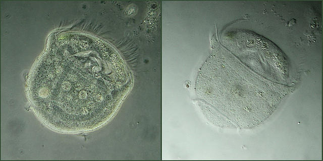
Image of Vorticella under phase contrast microscope:
One of the most distinct characteristics of Vorticella is that they have a cone-shaped body (bell-shaped). In the image above, it is possible to see fine hair-like structures on the upper part of the bell shaped organism. These fine hair-like structures are known as cilia and play an important role of sweeping food material into the peristome (wide opening surrounded by cilia).
On the second image, it's possible to see a slender stalk at the base of the organism. This plays an important role of attaching the organism to a substrate allowing the organism to float and feed on floating food material in the water. The unbranched stalk distinguishes Vorticella from the other ciliates.
One of its other characteristics is that the stalk contains myoneme which is a contractile fibril that makes it possible for the stalk to shorten and coil when stimulated. Therefore, when viewed under the microscope, it is possible to observe the stalk coiled up like a spring.
This is an image of a straight and spring-like stalk:
![Vorticella showing Springlike Stalk by Pearson Scott Foresman [Public domain], via Wikimedia Commons Vorticella showing Springlike Stalk by Pearson Scott Foresman [Public domain], via Wikimedia Commons](https://www.microscopemaster.com/images/Vorticella_springlikestalk.png) Vorticella showing Springlike Stalk by Pearson Scott Foresman [Public domain], via Wikimedia Commons
Vorticella showing Springlike Stalk by Pearson Scott Foresman [Public domain], via Wikimedia Commons
Measurements
Different species of Vorticella will vary in size. For Vorticella campanula, the bell shaped body has been shown to measure about 157 microns in length and 99 microns in width. The stalk on the other hand can vary considerably from about 53 microns to as long as 4150 microns.
Importance of Vorticella
In their environment, Vorticella species feed on a range of material and organisms including other smaller protozoa and bacteria. In their food system, this is important given that it helps control the population of these organisms in their environment.
This species are abundant in sewage treatment systems as well as aquatic farming ponds. As floating feeders, Vorticella species play an important role in such systems by consuming and breaking down organic matter in these environments.
In sewage treatment systems, they help in the breaking down of the organic matter, which and thus contributes in the treatment of the sewage along with other systems incorporated in the system.
More recently, the mechanism of these organisms is being incorporated into artificial engineering systems to help in the breakdown of various organic matter.
Return from Vorticella to Protozoa
Read about Protists
Return to Eukaryotes Main Page
See Ciliates Under the Microscope
Return to MicroscopeMaster Home
References
Ping Sun, John C. Clamp, Dapeng Xu, Bangqin Huang, Mann Kyoon Shin and Franziska Turner. (2018). An ITS-based phylogenetic framework for the genus Vorticella: finding the molecular and morphological gaps in a taxonomically difficult group.
Sangjin Ryu, Rachel E. Pepper, Moeto Nagai and Danielle C. France 4 (2016). Vorticella: A Protozoan for Bio-Inspired Engineering.
Toni Glymph. Wastewater Microbiology: A Handbook for Operators
Links
https://www.microscopyu.com/gallery-images/vorticella-protozoan-videos
https://biologywise.com/vorticella-reproduction
Find out how to advertise on MicroscopeMaster!


![Vorticella Species by Frank Fox (http://www.mikro-foto.de) [CC BY-SA 3.0 de (https://creativecommons.org/licenses/by-sa/3.0/de/deed.en)], via Wikimedia Commons Vorticella Species by Frank Fox (http://www.mikro-foto.de) [CC BY-SA 3.0 de (https://creativecommons.org/licenses/by-sa/3.0/de/deed.en)], via Wikimedia Commons](https://www.microscopemaster.com/images/Vorticella.jpg)


