Virus Vs Bacteria
Differences & Similarities in Size and Structure
Differences
Both bacteria and viruses are germs. As such, they are very tiny microbes/organisms/agents capable of causing diseases in human beings, animals as well as plants, etc. Although bacteria and viruses have a number of similarities (e.g. they are both microscopic etc), there are several differences that distinguish the two.
One of the biggest differences is that while bacteria are considered living things, viruses are not, or are at least placed somewhere between living and non-living. This particular difference between bacteria and viruses can be attributed to the structural differences between them as well as the manner in which they reproduce.
The following are structural differences between bacteria and viruses:
Structure
Cell wall, outer envelope, coating, membrane, and capsule - Although viruses are not regarded as cells, they, like bacteria, have an outer envelope that contains the inner contents of the particle. However, there are a number of differences between the outer envelope found in bacteria and those found in viruses.
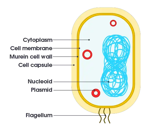
As compared to viruses, the majority of bacteria (about 90 percent of all bacteria) have a cell wall that consists of a peptidoglycan layer. Also known as murein, the peptidoglycan layer is a polysaccharide that consists of N-acetylmuramic acid and acetylglucosamine which alternate to form long chains. In addition to the two compounds, the layer also consists of a tetrapeptide (four amino acids) which cross-link the chains.
While the majority of bacteria have a cell wall that consists of peptidoglycan, the content of this layer varies between different types of bacteria which has allowed for bacteria to be divided into two main groups namely, Gram-positive bacteria (characterized by a thicker peptidoglycan layer) and Gram-negative bacteria (characterized by a thin peptidoglycan layer).
See more on Gram positive and Gram negative bacteria.
For bacteria with a cell wall, this structure performs a number of important functions that include maintaining the shape of the cell, protecting the cells from osmotic pressure, movement of substances in and out of the cell as well as in movement among others.
Cytoplasmic membrane /cell membrane - In addition to the cell wall, bacteria also have a cytoplasmic membrane located beneath the cell wall. Also known as the plasma membrane, the cytoplasmic membrane is a lipid bilayer that consists of about 40 percent phospholipid and about 60 percent proteins. It's characterized by a hydrophilic head (glycerol head) and a hydrophobic region that make up the tail part of the structure.
As compared to the cell wall, the cytoplasmic membrane is characterized by a fluid mosaic and is therefore not static. In addition to containing components of the cell, this membrane serves to regulate the movement of substances in and out of the cell.
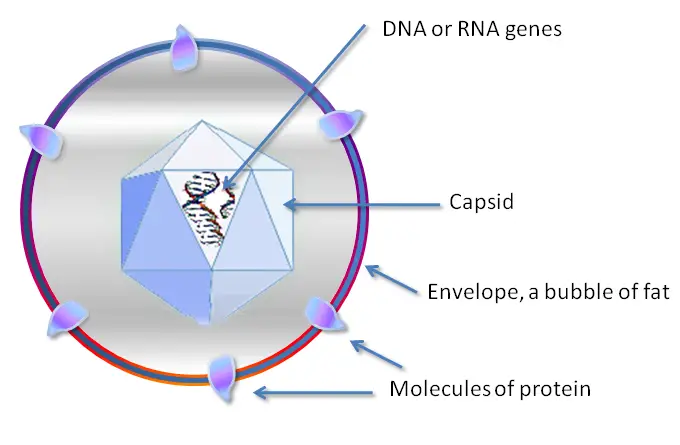
Capsid - As compared to bacteria, viruses consist of a capsid rather than a cytoplasmic membrane. Essentially, the capsid is a protein shell that encloses the nucleic acid content. As mentioned, the cytoplasmic membrane found in bacteria is a lipid bilayer that consists of phospholipids and proteins (however, it may also consist of several other components).
The capsid consists of subunits of proteins known as capsomeres. Depending on the virus, the capsid may consist of a single or double protein shell with a few structural protein species.
While the capsid, like a cell membrane, encloses the nucleic material among other contents, it's not primarily involved in the movement of substances in and out of the cell. As well, it's a robust complex which differentiates it from the more dynamic cytoplasmic membrane.
Viral envelope - In addition to the capsid, some of the viruses have an envelope surrounding the capsid. These viruses are known as enveloped viruses which distinguishes them from viruses without an envelope. For enveloped viruses, the envelope is a glycoprotein layer. However, the envelope also consists of two lipid layers with protein molecules (known as the lipoprotein bilayer).
Based on studies, their envelope has been shown to originate from infected cells (host cells). Here, new virus particles are wrapped in the envelope when they are released from the infected cell.
Over time, the viruses have been shown to add their own proteins to the envelope which allows them to survive and easily infect new cells With an envelope, they can easily fuse with the cell membrane of the host cell which allows them to easily enter the cell.
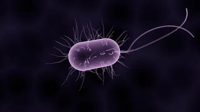
Other components - there are a number of other components that can be found on the capsid, envelope, membrane, and cell wall that distinguish bacteria from viruses. For instance, the surface of some bacteria consists of flagella, pili, or both.
For the most part, these structures contribute to motility, adhesion, or attachment of the cell which may contribute to their pathogenicity. For instance, using pili, some bacteria are able to attach to the cell of the host and invade the cell. However, for free-living bacteria, flagella may be used to swim in aquatic environments.
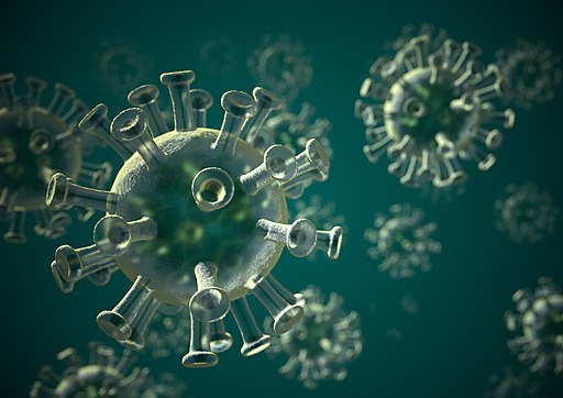
Viruses, on the other hand, do not have flagella or pili. However, they may possess a tail sheath among other attachment proteins that allow them to attach to the surface proteins or structure (e.g. viral spikes) of the host cell before they can invade the cell. For some bacteria, an outer capsule may be present (known as the outer envelope).
As compared to the cell wall or the cytoplasmic membrane, this capsule (also referred to as a slime layer) is a polysaccharide layer that has been shown to help the organism develop resistance to various drugs. In addition, it protects the cell from desiccation thus allowing some bacteria to survive under harsh environmental conditions. Viruses, on the other hand, do not possess this structure.
Cellular Components
In general, the structure of a virus is very simple, mostly consisting of the nucleic acid, the capsid, as well as the outer envelope for some viruses. Viruses also may contain an attachment structure that allows them to attach to the surface of the host cell.
As compared to viruses, bacteria contain a few organelles that perform different functions which allow the organisms to obtain energy from their surrounding, grow, and reproduce. For this reason, they are considered living organisms as they have in place various cellular mechanisms that allow them to grow and reproduce.
The following are some of the cellular components of bacteria and viruses:
Genetic material - Both bacteria and viruses have genetic material (nucleic acid). In bacterial cells, the genetic material is contained in a chromosome which is a strand of DNA. While they do not have a nucleus, the genetic material of these organisms is contained in a region generally known as the nucleoid.
In addition to the nucleoid, bacteria also have a plasmid, which is an extrachromosomal molecule of DNA. For the most part, the plasmid exists as a circular, double-stranded DNA molecule that can replicate on its own.
Viruses contain nucleic acid in the form of DNA or RNA. Unlike bacteria, however, viruses do not have a plasmid (extrachromosomal DNA).
Ribosome - In addition to the genetic material, bacteria also have ribosomes (composed of two subunits) which are nucleoprotein particles involved in mRNA translation for protein synthesis.
Unlike the ribosome found in eukaryotic cells (on the rough E.R), bacterial ribosomes are not bound to any cellular structures/organelles. As such, they are freely distributed in the cytoplasm. Unlike bacteria, viruses do not have ribosomes and thus rely on organelles or the host cell for protein synthesis.
Other structures of the cell - As already mentioned, different types of bacteria contain a number of additional structures/organelles including flagella, pili, and fimbriae which are primarily involved in attachment or movement.
Bacterial cells also contain the cytoplasm/protoplasm in which metabolic activities and replication take place. The cytoplasm contains a variety of substances including gases, waste material, and water among others.
In viruses, the cytoplasm may not be present. While viruses do not have many of the structures/organelles present in bacteria, they have attachment structures such as glycoprotein spikes and tail fibers, etc that allow them to attach onto the surface of the host cell.
However, in general, they have a simple structure that is aimed at ensuring the successful transfer of their genetic material into the host cell for transcription and reproduction purposes.
Size
The other significant difference between bacteria and viruses is with regard to size. Although research studies have revealed that some viruses are very large in size (e.g. the Mimivirus is about 750nm in diameter), viruses are generally very small when compared to bacteria.
Some of the smallest viruses may range from 20 to 30 nm in diameter (e.g. members of the families Parvoviridae and Picornaviridae) with the majority of viruses ranging between 10 and 300nm in diameter.
Bacteria are relatively larger, ranging between 0.2 and 2.0 micrometer in diameter (the largest bacteria has been shown to be about 750um/0.75mm in diameter). Therefore, in general, smaller viruses can be over 200 times smaller when compared to some bacteria. Because of their small size, some viruses are also capable of infecting bacteria.
These viruses are commonly known as bacteriophages. By injecting their nucleic material into the bacteria, they are able to take over the reproductive machinery of the cell and make many copies of themselves.
Activity
Because of the differences in their general structures, bacteria and viruses vary significantly when it comes to various activities.
The following are some of the activities related to reproduction and disease-causing mechanisms:
Replication - Replication refers to the process through which the DNA molecule is copied for the purposes of producing two identical copies. In bacteria, the process starts at the origin of replication (on the DNA strand). Here, the DNA strand is first unwound (by the enzyme Helicase).
Another enzyme known as DNA Gyrase ensures that the double-stranded area located behind the replication site do not supercoil. Once the two strands are ready (when the replication fork is stable), DNA polymerase serves to catalyze the addition to new nucleotides onto the growing daughter strands.
Ultimately, this results in the production of two new DNA strands that coil to form a helix before the cell divides into two. This process ensures that the new daughter cells have the same genetic material as the parent cell.
Given that viruses do not have the machinery required for replication, they have to rely on the machinery of the host cell. For this reason, replication can only take place when they invade the cell of the host. This may be achieved through endocytosis or using surface molecules on the host cell.
Following the entry process, the virus has to uncoat in order to release the genetic material. Using the machinery of the host cell, the nuclei material is able to replicate and produce a new strand. Here, however, it's worth noting that there are various types of viruses with different genetic material. Therefore, the manner in which transcription takes place will vary between different viruses.
Regardless, they all depend on the machinery of the host cell for this process to be successful. Following successful replication, the virions are assembled as newly formed strands of nucleic acid and structural proteins, to form a nucleocapsid. The newly formed viral particles then spread to new cells as the process continues.
Disease-causing mechanism - While some bacteria are intracellular pathogens, not all bacteria depend on the host cell to develop. For this reason, some parasitic bacteria do not have to invade cells for diseases to occur. For instance, some bacteria, like Clostridium botulinum, cause disease by releasing toxins that affect cells and various tissues.
Viruses are obligate intracellular parasites and therefore cannot reproduce outside the cell. Once they enter and start reproducing inside the cell, they not only affect normal cell processes but may also rupture the cell as they increase in numbers. In the process, they cause significant damage to cells. This is evident in respiratory infections such as mild flu and tonsilitis.
* Not all bacteria cause diseases.
Translation – Translation refers to the process through which information in mRNA is decoded for protein synthesis. As is the case with transcription, the components of the host cell are used for the purposes of making viral components.
While the information is contained in the mRNA of the virus, they do not have the necessary machinery to translate this information into protein. For this reason, the host cell is instructed to produce viral components using information contained in the mRNA of the virus.
Given that bacteria have ribosomes as well as the necessary enzymes, information from the mRNA is immediately decoded and used to produce proteins in the cytoplasm.
Virus Vs Bacteria: Similarities
Although they have a number of differences, bacteria and viruses have a number of similarities.
These include:
Lack membrane-bound organelles - While bacteria have a few organelles involves in metabolism and reproduction, they, like viruses, do not have membrane-bound organelles. In viruses, the nucleic acid is not contained in a nucleus as is the case with eukaryotic cells. Here, however, it's also worth noting that unlike bacteria, viruses are not really considered to be cells.
Both microscopic - Both bacteria and viruses are microscopic and therefore too small to be seen with the naked eye. While they vary in size, they are both very small and therefore a microscope has to be used to study their general morphology and structure.
Diseases - While some bacteria are actually beneficial to human beings others are free-living bacteria and viruses that can cause diseases in human beings, animals, as well as plants etc. Following entry into the body of the host, they can both directly or indirectly affect various normal processes and thereby cause disease.
Both carry genetic material - While viruses are not necessarily considered living organisms, they, like bacteria, carry genetic material that is replicated and used to form new individuals. However, the manner in which this is achieved between the two is different as mentioned above.
See also: Bacteriology, Virology
List of Diseases caused by Bacteria
Return to Bacteria under a Microscope
Return to Bacteria - Size, Shape and Arrangement
Return from Virus Vs Bacteria to MicroscopeMaster home
References
Hans R. Gelderblom. (1996). Structure and Classification of Viruses. Medical Microbiology. 4th edition.
Matthew D. Moore and Lee-Ann Jaykus. (2018). Virus–Bacteria Interactions: Implications and
Potential for the Applied and Agricultural Sciences.
Scott M. Hammer. Viral Replication.
Links
https://open.oregonstate.education/generalmicrobiology/chapter/bacteria-surface-structures/
https://www.cedars-sinai.org/blog/germs-viruses-bacteria-fungi.html
Find out how to advertise on MicroscopeMaster!


