Streptococcus Bacteria
Classification, Shape, Infection & Gram Stain
Overview
Streptococcus bacteria is Gram-positive and are generally spherical in shape. They are commonly found in the mucous membrane of the mouth and respiratory tract etc where they have been associated with a number of diseases and infections including sepsis, pneumonia, and pharyngitis.
Classification
Kingdom: Bacteria - As members of the kingdom bacteria, Streptococci are prokaryotes and thus lack membrane-bound organelles. They also contain a peptidoglycan layer in their cell wall and can be found in various ecological habitats.
Subkingdom: Posibacteria - Streptococcus bacteria are also classified under the subkingdom Posibacteria which largely consists of Gram-positive bacteria
Phylum: Firmicutes - Firmicutes is a phylum of the kingdom bacteria that consists of Gram-positive bacteria (a few members like Selenomonas may have characteristics associated with Gram-negative bacteria). Along with a number of other bacteria, members of this phylum are part of the healthy gut microbiota composition.
Class: Bacilli - Bacilli is a class of bacteria characterized by low G+C content. This group consists of Gram-positive bacteria and is divided into two main orders that include Bacillales and Lactobacillales. While the name Bacilli suggests that members of this group are rod-shaped, it's worth noting that not all organisms in the class are rod-shaped (e.g. streptococci and staphylococci).
Order: Lactobacillales - Lactobacillales is an order under the class Bacilli that consists of both rod-shaped and spherical bacteria. They are Gram-positive bacteria. They are characterized by low G+C content and are often referred to as lactic acid bacteria
Family: Streptococcaceae - Members of the family Streptococcaceae include Streptococcus bacteria and Lactococcus among others. They are Gram-positive, spherical bacteria that grow in chains or pairs. They have also been shown to be facultative anaerobes that ferment carbohydrates.
Genus: Streptococcus: Characteristics of this group of bacteria are discussed below in detail.
Some examples of Streptococcius bacteria include:
- Streptococcus pneumoniae
- Streptococcus mutans
- Streptococcus bovis
- Streptococcus salivarius
- Streptococcus oralis
- Streptococcus canis
Shape
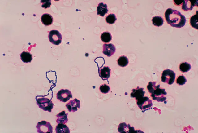
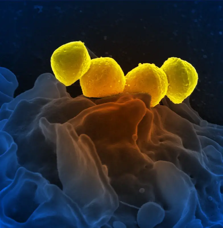
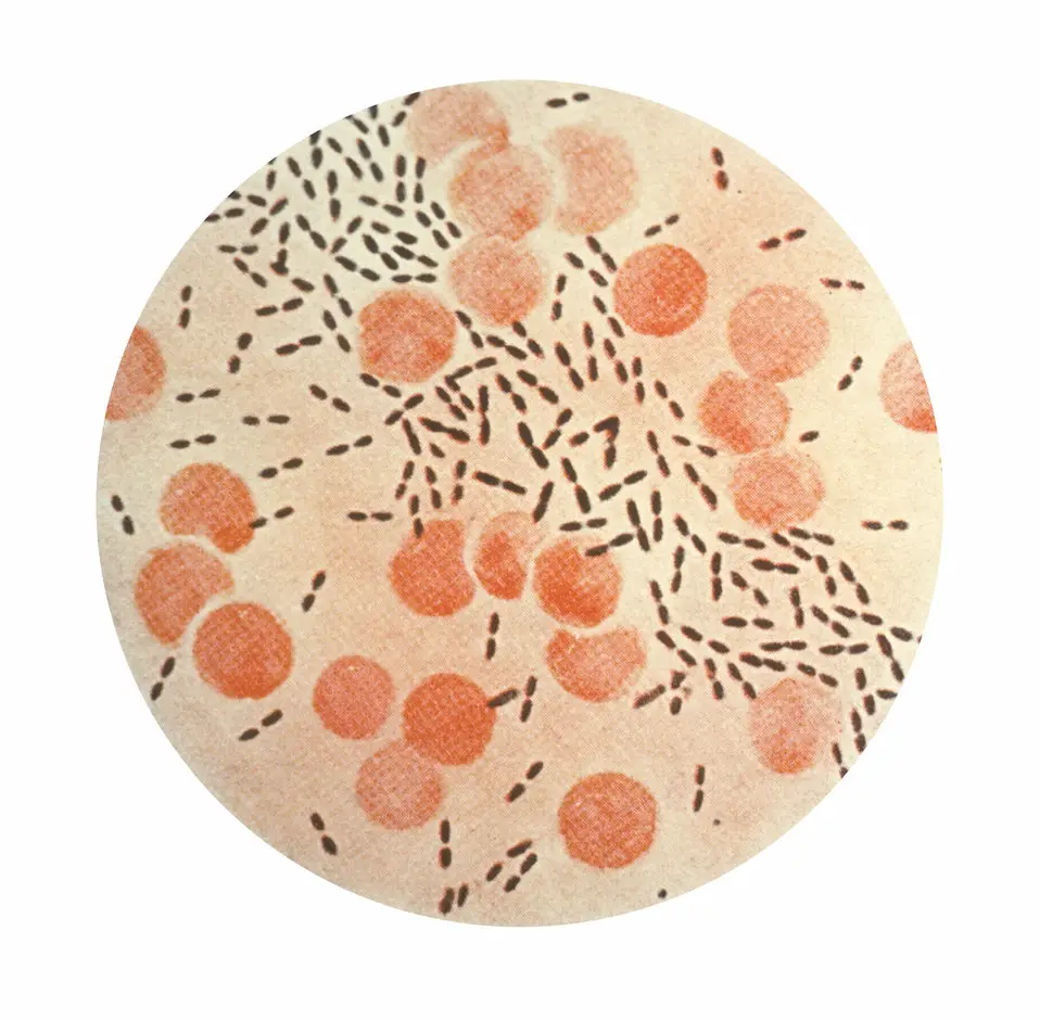
Like many other bacteria, Streptococcus bacteria are small in size, ranging from 0.5 to 2.0 micrometers in diameter. Here, however, the majority of species are less than 2um in size. With regards to shape, Streptococci may appear spherical or ovoid in shape. When viewed under the microscope, Streptococci occur in chains (resembling a string of beads) or in pairs.
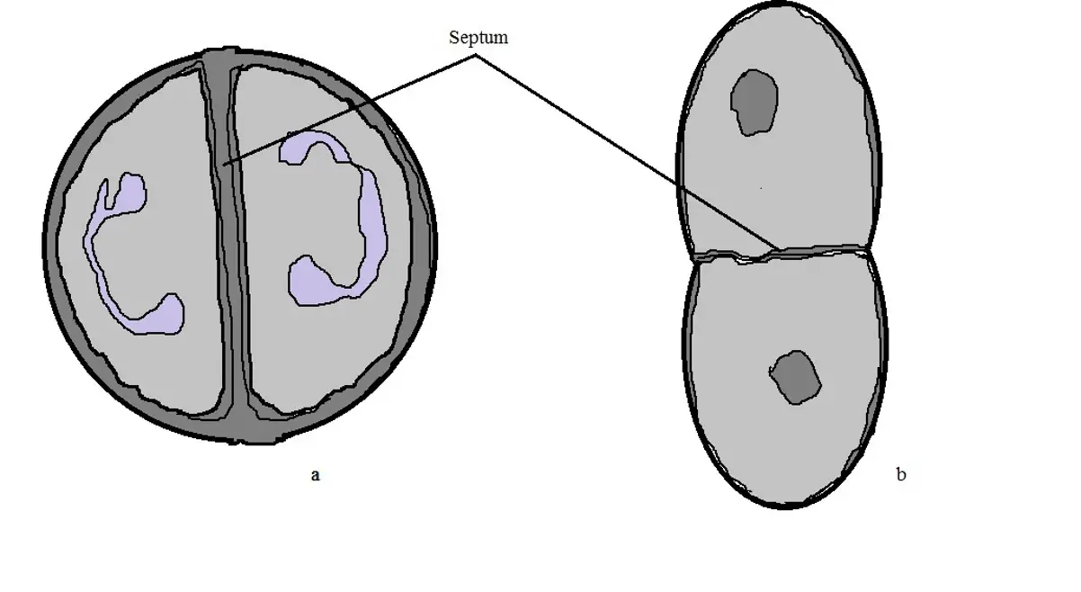
As is the case with other bacteria, the general shape of Streptococci is maintained by the manner in which they propagate. In the case of Streptococcus pneumoniae, the synthesis of the peptidoglycan during propagation is suggested to occur through peripheral and septal systems. This is different from the more spherical species where only the septal system is involved.
In the case of Streptococcus pneumoniae, division is characterized by inward growth of the cross wall located at the cell equator. At this stage of division, microscopic studies have revealed a wall band, also known as the equatorial ring at the region of division, followed by the formation of two new equatorial rings as new peptidoglycan is gradually inserted between these rings. This allows for peripheral growth to continue until the internal hemisphere formed reach the size of the old ones.
The end of peripheral growth is followed by septal synthesis of the peptidoglycan. Here, peptidoglycan hydrolases are involved in splitting the septum thereby separating the daughter cells.
Unlike micrococci and deinococci, which are truly round cells, streptococcus bacteria may appear elongated (ovoid in shape). According to studies, this is also due to the fact that their division is along a successively parallel plane (which is perpendicular to their long axis).
This is different when compared to such truly round cocci such as micrococci and staphylococci where division may occur in two or three alternating perpendicular planes. Therefore, the cell wall, along with the plane of division have a role to play in the general shape of bacterial cells.
* Depending on the species, the length of Streptococcus bacteria chains has been shown to play an important role in virulence (e.g. studies have shown bacterial invasion in Streptococcus pneumoniae to be directly related to the chain length).
* A number of factors influence the length and nature of the chain (bacterial chain) formed. For instance, whereas normal short/long chains are formed in vivo, cultures have been shown to produce clustered chains.
Streptococcus Bacterial Infection
With the exception of a few species (e.g. lactic acid group that is commonly associated with plant environment and dairy), the majority of Streptococcus bacteria occupy various parts of the human and animal bodies.
Based on a number of studies, the nutritional needs of these organisms are met by their hosts. For this reason, they tend to thrive through their relationship with animal hosts. Because of their relationship with some animals and human beings, Streptococci are widely distributed across the world (wherever their hosts exist).
In their relationship with their respective hosts, Streptococci either exist as commensals or as parasites. As commensals, they can live in harmony with the host (where they derive nutrition from the host without causing any harm).
As parasites, they cause harm to the host by causing diseases. Depending on the organism they infect and host specificity, Streptococci bacteria can be divided into several groups.
The pyogenic group as well as species that are often found in the intestine are commonly found in homeothermic hosts. Here, however, some species are only found in specific hosts and are known as host-specific bacteria. For instance, whereas Streptococcus pyogenes are commonly found in human beings, Streptococcus causes infections in horses.
Other species can infect a number of animals, as well as human beings and can be found in given parts of their hosts. Such species as Streptococcus equinus and Streptococcus bovis, of which belong to the subthermophilic group, are intestinal dwellers and can, therefore, be found in the intestine of several hosts.
While anaerobic Streptococcus bacteria have been shown to be part of the normal flora in the mouth, genital tract, skin, and genital tract, etc, a number of species are responsible for various infections that range from mild to severe.
They are divided into two main groups that include:
Alpha Hemolytic Streptococcus
Like the other members of the genus Streptococcus, this group consists of Gram-positive bacteria that are non-motile and normally form short or long chains. They are anaerobic and cause oxidation of iron in hemoglobin.
Members of this group produce hydrogen peroxide which causes hemolysis, converting hemoglobin into methemoglobin. Species found in this group are particularly common and live naturally in human beings without causing harm. However, some of the species are responsible for infections in human beings.
Some of the species responsible for human infections include:
Streptococcus pneumoniae
Streptococcus pneumoniae is normally found in the upper respiratory tract. According to statistics, it's estimated that between 27 and 65 percent of children and less than 10 percent of adults are carriers of the bacteria.
Although it normally exists as a commensal in human beings, it's an opportunistic pathogen and causes infections when it spreads to other parts of the respiratory system or enters the bloodstream and spreads to other parts of the body.
Generally, transmission of Streptococcus pneumoniae occurs through direct contact between two individuals through respiratory droplets. Given that children tend to play close together, this is one of the reasons why the infection rate is higher among children than adults.
Once the bacteria has been transmitted from an infected individual/carrier to another person, a number of factors allow the organism to colonize and persist in the mucosal surface of the respiratory tract. These include the expression of two important enzymes by the bacteria that modify the peptidoglycan thus rendering it resistant to lytic activities of the lysosome, adhesion to the local cells and tissue as well as the ability to evade the clearance system of the mucosal surface.
In the event that the bacteria are able to invade these tissue and spread or enter the bloodstream, they then spread to other parts of the body (e.g. ear, brain, etc) where it causes infections.
While it causes pneumonia in the lungs, Streptococcus pneumoniae is responsible for meningitis when it invades the membrane covering the brain and spinal cord and bacteremia when it enters the bloodstream.
* Children with a poor immunity and the elderly are at higher risk of these infections.
Streptococcus Viridans
Like Streptococcus pneumoniae, Viridans streptococci are also an Alpha-hemolytic Streptococcus. They are also part of the normal microbial flora and are abundant in the oral cavity in human beings. However, they can also be found in other parts of the body including parts of the gastrointestinal tract, the upper respiratory tract as well as the genital tract in females.
Like Streptococcus pneumoniae, they exist normally as commensals but can invade tissue in these parts of the body and cause diseases. They are opportunistic pathogens and cause disease in immunocompromised individuals or when the mucosa in which they reside is disrupted.
In the event of trauma in the mucosa (e.g. oral mucosa), Viridans get an opportunity to enter the bloodstream where they can cause bacteremia. As well, they can be transported to the heart where they can cause Endocarditis.
Here, the bacteria are transported to the heart where they may adhere to the surface of the heart valves (particularly in cases where these valves had been previously damaged). In cases where the bacteria start to multiply on these surfaces, studies have shown them to be covered by fibrin and platelets resulting in the formation of macroscopic verrucous lesions, abscesses, pericarditis as well as peripheral emboli, etc.
* According to statistics, this particular bacteria is responsible for between 45 and 80 percent of cases of native valve endocarditis in human beings.
Some of the clinical manifestations of Viridans Streptococci infections include:
· Fever - This is the first sign of Viridans Streptococci infections and is indicative of bacteremia. It may occur several days after the infection and tends to persist for days.
· Toxic shock-like syndrome - Some patients exhibit toxic shock-like syndrome in cases where the infection persists in the blood.
· Rashes in the palm - Some patients may exhibit desquamation of the palms.
· Chills
· Flushing Pharyngitis
· General weakness
Treatment of Alpha-hemolytic infections
- Vancomycin
- Quinupristin-Dalfopristin
- Telithromycin
Beta Hemolytic Streptococci (β-Hemolytic streptococci)
The second group of Streptococci bacteria responsible for various human infections/diseases is known as beta (β)-haemolytic Streptococci. Like Alpha Hemolytic Streptococcus, members of this group are also Gram-positive bacteria that grow in pairs or form chains. Members of this group cause lysis of red cells using hemolysins O and S.
Members of this group are divided into two main groups that include:
Group A Streptococci (Streptococcus pyogenes)
Streptococcus pyogenes is a Gram-positive bacteria that is only found in the skin and mucous membrane of human beings. It has also been shown to colonize the vaginal and rectal regions.
Globally, the bacteria are responsible for a variety of infections some of which can be life-threatening. Given that it's commonly found in the skin and mucous membrane, Streptococcus pyogenes is transmitted through respiratory droplets as well as direct skin contact. According to a number of studies, there have been cases where disease outbreaks resulted from good-borne Group A Streptococci.
Following the initial transmission, the bacteria adheres to the cells and extracellular matrix proteins at the site of colonization using Lipoteichoic acid and a number of other proteins. This allows for adherence to the epithelial cells and the consequent internalization of the bacteria.
Being a human-adapted pathogen, Streptococcus pyogenes is able to evade the actions of innate immune defenses by producing a variety of surface-bound virulence factors such as antimicrobial peptides.
Some of the infections associated with the bacteria include:
Cellulitis - This is an infection of the subcutaneous tissues. For the most part, it's transmitted by individuals with an active infection such as athlete’s foot providing a portal of entry for the bacteria. Cellulitis is normally characterized by skin redness and inflammation as well as pain and swelling.
Bacteremia - Bacteremia occurs when the bacteria enter the bloodstream. This infection is characterized by vomiting as well as a high fever. Bacteremia may occur if cellulitis is not treated.
Necrotizing fasciitis - This is a serious infection that affects the subcutaneous layer, deep tissue, and fascia. Being an invasive bacterial infection characterized by rapidly spreading necrosis, Necrotizing fasciitis has a high mortality rate.
Some of the other infections associated with the bacteria include:
Myositis - An infection of the muscles with very high fatality rates
Lymphangitis - Lymphangitis is often associated with cellulitis. However, it may also occur following a minor skin infection
Vulvovaginitis - This is an infection of the vulvar and vaginal area in females and is characterized by intense pruritus
Streptococcal toxic shock syndrome (STSS) - STSS is commonly associated with necrotizing fasciitis, pneumonia, and post-partum infection, etc. As such, it's likely to occur when the bacteria gains entry through damaged skin or mucus membrane. It's characterized by chills, vomiting, and myalgia. if not treated early enough, then organ failure and death may result.
Pneumonia – Infection of lung tissue
* There are many other infections associated with Group A Streptococci.
Group B Streptococcus (Group B Strep Infection)
Group B Streptococcus (S. agalactiae) normally resides in the digestive tract and female genitals as a commensal. Therefore, under normal conditions, it does not cause harm. It's beta-hemolytic and thus causes complete lysis of the red cells (using Streptolysin).
S. agalactiae is responsible for sepsis and meningitis in newborns (neonates) as well as infections in elderly patients. For the most part, transmission from the mother to the child occurs through the amniotic fluid of the mucus membrane. Sexual contact between adults has been shown to play a role in the transmission of the bacteria (fecal-oral transmission).
Following a successful transmission, the bacteria may result in the following infections:
- Cellulitis, foot infection, or decubitus ulcers
- Endocarditis
- Osteomyelitis
- Pneumonia
- Meningitis
- Urinary tract infections
Gram Stain
In addition to Optochin test, bile solubility test, and catalase test, Gram staining is one of the methods used to determine the presence of Streptococcus bacteria in a sample. In this section, S. pneumoniae will be used as the representative of the group.
Requirements
For Gram staining, the following material will be required:
- Sample - A sputum sample of patients with pneumonia may be used
- Compound microscope
- Reagents (Gram stain reagents)
- Glass slide
- A burner
- Water
- Oil immersion
Procedure
· Using a sterilized wire-loop or a sterilized applicator stick, create a smear of the sample at the center of a clean glass slide
· Fix the specimen by passing the slide over a burner - take care not to overheat
· Allow the slide to cool and then cover with the primary stain (crystal violet) for about 1 minute
· Rinse the slide with water then add the mordant for one minute (iodine solution) - Adding the mordant to the sample creates a complex between the primary stain and the iodine
· Wash the slide with running water
· Cover the slide with the decolorizer (ethyl alcohol or acetone) for a few seconds - You can do this by adding the decolorizer dropwise
· Add the counterstain (safranin) and allow the slide to stand for about 1 minute
· Rinse the excess stain with water and allow the slide to air-dry
· Mount the slide on the microscope and observe under oil immersion - 100x
Observation
When viewed under the microscope at 100x, Streptococcus pneumoniae will appear as purple single cells or in pairs. However, it's possible to identify chains consisting of between 4 and 6 cells.
As compared to Streptococcus pneumoniae, other Streptococci may have longer cells consisting of more than 10 cells. When compared to Streptococcus pneumoniae, Streptococcus agalactiae appear to have longer chains consisting of 10 or more cells.
How do Bacteria cause Disease?
Learn more about Gram positive and Gram negative Bacteria
Return to learning more about Bacteria like Shape, Size and Arrangement
Return from Streptococci Bacteria to MicroscopeMaster home
References
Allan R. Tunkel and Kent A. Sepkowitz. (2002). Infections Caused by Viridans Streptococci in Patients with Neutropenia.
Jeffrey N. Weiser, Daniela M. Ferreira, and James C. Paton. (2018). Streptococcus pneumoniae: transmission, colonization and invasion.
J. Orvin Mundt. (1983). The Ecology of the Streptococci.
Mark J. Walker et al. (2014). Disease Manifestations and Pathogenic Mechanisms of Group A Streptococcus.
Maria Jevitz Patterson. Streptococcus.
Links
Find out how to advertise on MicroscopeMaster!


