Staphylococcus Bacteria
Examples, Classification and Characteristics
Overview
Staphylococcus bacteria, identified as the cause of various pyogenic infections in man in 1880 (by Sir Alexander Ogston), are Gram-positive characterized by irregular clusters. They are widely distributed in nature and can be found on the skin and mucous membranes (nasopharynx and gastrointestinal tract) of various animals and birds.
The genus is divided into coagulase-positive staphylococci which are pathogenic and the coagulase-negative staphylococci (e.g. Staphylococcus epidermidis) which are common commensals on the skin. They can behave as opportunistic pathogens under certain conditions and cause infections. As extracellular bacteria, Staphylococci bacteria are very hardy and thus capable of surviving for a long period of time on various surfaces.
Common examples of Staphylococcus bacteria include:
- Staphylococcus aureus
- Staphylococcus epidermidis
- Staphylococcus saprophyticus
- Staphylococcus haemolyticus
- Staphylococcus intermedius
- Staphylococcus warneri
* The name Staphylococcus is derived from the Greek words "staphyle" which means grapes and "coccus" which means a grain or berry.
Classification of Staphylococcus
Domain: Bacteria - Single-celled prokaryotes widely distributed in every environment across the world. They exist as parasites or as free-living organisms with some of the species being beneficial to man.
Phylum: Firmicutes - The phylum Firmicutes consists of many Gram-positive bacteria with a few species exhibiting Gram-negative properties. They also produce inactive spores (some species) that survive various environmental stresses
Class: Bacilli - The class Bacilli consists of Gram-positive bacteria with low G+C - less than 50 percent of guanine and cytosine in their DNA. While the majority of species have a rod-shape appearance, their shape can range from branching thin filamentous rods to coccobacilli depending on the species.
Order: Bacillales - The order Bacillales consists of Gram-positive bacteria most of which have a small rod shape. In a majority of the representative genera, the majority of species have also been shown to form endospores.
Family: Staphylococcaceae - Members of this family are Gram-positive bacteria. They are commonly found on the skin, intestine, the respiratory system, and oral cavity and are capable of causing suppurative lesions and septicemia under certain conditions.
Genus: Staphylococcus (Characteristics of the genus Staphylococcus are discussed below)
Distribution and Ecology
Members of the genus Staphylococcus are distributed worldwide and can be found wherever human beings and other warm-blooded animals exist. Early on, researchers noticed that Staphylococcus bacteria are often found in association with other organisms (particularly warm-blooded organisms).
This led to the conclusion that mammalian skin acts as the major habitat of these organisms. However, some of these organisms have also been identified in other regions of the body including the mouth, intestinal tract, and throat among others.
In these regions of the body, staphylococcus occur less frequently compared to the skin where they exist in high numbers. Apart from mammals, Staphylococcus species have also been isolated from birds.
While Staphylococcus species exist more frequently on exposed skin, they can easily be washed away as transients. For this reason, they exist in few numbers in clean skin. Here, however, it's worth noting that members of the genus are classified into two major groups (transients and residents) based on their presence in various regions of the body of mammals.
Transient species are those that come from extraneous sources and land on the skin of mammals. Those forms are temporary residents given that they can easily be removed by washing the skin. Residents, on the other hand, are indigenous and tend to persist for extended periods of time.
As compared to resident Staphylococcus, transient species have also been shown to be present in low numbers and do not reproduce rapidly. During a routine surgical hand scrub, the transient forms are completely destroyed while residents are reduced in number.
General Morphology and Cell Structure
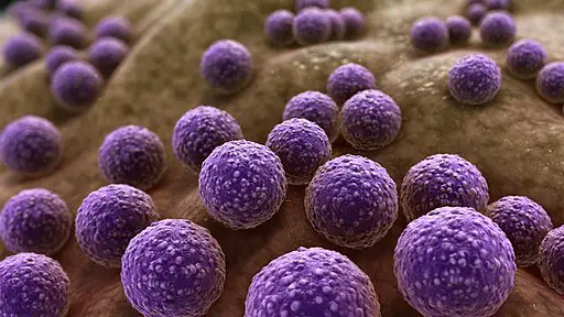
As the name suggests, Staphylococcus bacteria have a spherical shape (cocci). Unlike other cocci bacteria that may exist as single cells or in pairs etc, Staphylococcus occurs in clusters and thus resemble grape-like clusters.
In these clusters, individual cells may vary in size. However, this variation has also been detected between different species with the size ranging from 0.5 to 1mm in diameter.
When viewed under the microscope, some species have been shown to produce dwarf colonies that have a relatively smooth and glossy appearance. Generally, staphylococcus bacteria are non-motile and therefore lack structures (e.g. cilia or flagella) required for movement.
* The Grape-like cluster of Staphylococcus is the result of a single coccus dividing in more than one plane.
* Some of the species (e.g. staphylococcus aureus) produce pigments that cause them to appear yellowish/golden in color when viewed under the .
Cell Wall
For the majority of bacteria, the cell is surrounded by a cell wall. This cell wall is composed of peptidoglycan and teichoic acid in Staphylococcus (which are also used to distinguish different Staphylococcus species).
While peptidoglycan type L-Lys-GlY5-6 is found in several Staphylococci (Staphylococcus aureus, Staphylococcus hycus, and Staphylococcus cohnii etc), the composition of teichoic acid between these species varies which makes it possible to distinguish between them (e.g. Staphylococcus aureus contains N-acetylglucosaminyl ribitol teichoic acid while Staphylococcus simulans contains glycerol teichoic acid).
Current. three main components have been identified in the cell wall of Staphylococci bacteria.
These include:
- Wall-associated surface proteins
- Murein
- Teichoic acids
The murein, which consists of glycan strands cross-linked by peptide furnishing, is continuous and covers the entire cell. Being a macromolecular sacculus, murein is one of the main distinctive features of Staphylococci bacteria with a high degree of cross-linking.
Teichoic acid, on the other hand, is first produced as corresponding biosynthetic precursors that resemble the precursors involved in murein biosynthesis. Along with surface-associated proteins, studies have shown the precursors of teichoic acid to attach to the murein during its assembly.
* Under the electron microscope, the cell wall of Staphylococcus aureus has been shown to be relatively thick, ranging from 20 to 40nm. This is characteristic of Gram-positive bacteria that contain a thicker peptidoglycan compared to that of Gram-negative bacteria.
* The murein makes up the peptidoglycan of the cell wall and thus giving it structural strength.
* The teichoic acid, which is also an important component of the cell wall, contributes to bacterial physiology and thus contributes to host interaction, resistance to antimicrobial substances as well as enhancing virulence.
* Surface proteins are covalently attached to the peptidoglycan and serve a range of functions including promoting pathogenesis.
Reproduction and life cycle (Staphylococcus aureus)
Like many other bacteria, Staphylococci bacteria divide through binary fission. While this process produces two daughter cells, they do remain attached to one another which eventually results in clustering. Based on various studies, this process in Staphylococcus aureus has been shown to be under the control of GpsB (an essential protein).
Here, GpsB is suggested to interact with FtsZ (an important component of cell division machinery in bacteria) increasing its GTPase activity. This contributes to the constriction of the cell envelope ultimately causing the cell to divide into two.
During cell division (through binary fission), the peptidoglycan also increases in size as the cell elongates (the cell wall grows outwards from the FtsZ ring). In the process, the backbone of the peptidoglycan is severed which necessitates the synthesis of new cell wall material.
This is made possible by autolysins (enzymes) which hydrolyze glycosidic bonds that are then linked to N-acetylglucosamine and N-acetylmuramic acid with the gaps being filled by various material of the cell wall. Ultimately, this allows each of the newly formed cells to have its own cell wall consisting of peptidoglycan.
Nutritional Adaptations
Staphylococci species have been shown to circulate between various nutrient-limiting environments including terrestrial habitats and host species among others. In the event of limited nutrients, they persist on given environs for a long time which allows them to survive until conditions improve.
For instance, when Staphylococcus aureus are grown in a glucose-limiting media, a small remnant of the bacteria (which become smaller in size) are able to live for a long period of time with the ability to survive stressful conditions. This is one of the reasons Staphylococci species are very problematic in clinical settings where they infect medical devices and various surfaces etc.
* On the skin, commensals such as coagulase-negative staphylococci are beneficial given that they promote/boost the competence of the cutaneous immunity by enhancing the innate and adaptive immune cell network on the skin. This, in turn, controls the other normal flora on the skin.
Pathogenesis
Although a good number of Staphylococci bacteria exist as commensals on various regions of the body, they can cause various infections some of which can be fatal if untreated. Depending on the conditions, these infections may be a result of a mechanical breach in the skin/mucosal barriers or as a result of toxins produced by the bacteria.
Given that they frequently present on the skin as well as various other sites in the body, Staphylococcus species, e.g. S. aureus can take advantage of wounds or intravascular catheters, etc to cause diseases. Whenever they get an opportunity to invade body tissues, they colonize and invade before spreading to other parts of the body through circulation.
In cases where they penetrate the skin and reach the internal body tissues, staphylococcus use their surface proteins (e.g. collagen-binding protein to attach to the surfaces of these tissues). This promotes adherence as they produce enzymes that allow them to obtain nutrients. As they grow and increase in numbers, they start spreading to adjacent tissue.
At times, they enter the bloodstream causing bacteremia. Apart from bloodstream infections, S. aureus is also responsible for bone and joint infections as well as pneumonia in some patients.
Apart from invasive infections, Staphylococcus can also cause diseases by releasing toxins. Some of the toxins commonly released by S. aureus are hemolysin and leukotoxin. These toxins can cause toxic shock syndrome and scalded skin syndrome as well as food poisoning.
Here, these diseases have been shown to occur when the toxins bind to antigen-presenting cell molecules. These toxins are capable of causing biological damage to membranes of the cell thus causing cell death. In some cases, they have also been shown to lyse neutrophils once they are ingested, an action that protects the bacteria from host innate immunity.
Some of the other forms of infections caused by Staphylococci bacteria include:
- Superficial skin lesions as well as localized abscesses
- Endocarditis
- Urinary tract infections
Culture and Microscopy
Using culture media, it is possible to isolate and maintain pure cultures of a given organism. This also makes it possible to determine whether a given microorganism of interest is present in a sample.
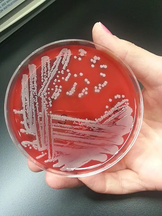
Blood agar is commonly used as a differential and enriched medium to culture Staphylococci bacteria. Here, blood is used as the enrichment ingredient for cultivating fastidious organisms.
If present in the sample, various Staphylococcus will appear differently. For instance, S. aureus exhibits a light or golden yellow color which is indicative of pigmentation. S. epidermidis appear white in color while S. saprophyticus exhibits a bright yellow or white color.
Gram Stain
Gram staining technique is used to identify the different types of bacteria using Gram stain.
Requirements
- Sample
- Glass slide
- Crystal violet
- Safranin
- Iodine solution
- Microscope - Compound microscope
- Burner
- Water
Procedure
On a clean glass slide, create a thin smear of the sample - This can simply be achieved by placing a drop of suspended culture on a slide using an inoculation loop and smearing to create a thin layer.
Allow the sample to air dry and then fix by passing over a gentle flame - Move the slide several times over the flame to avoid localized overheating.
Pour crystal violet stain on the culture and allow to stand for about 30 seconds to 1 minute.
Pour off the stain and rinse with water
Add iodine solution making sure to cover the fixed sample and allow to stand for between 30 seconds and 1 minute.
Pour off the iodine solution and rinse with water.
Add a few drops of the alcohol decolorizer and rinse with water for about 5 minutes.
Counter-stain the sample using safranin or basic fuchsin for about 1 minute and then wash with water.
Remove excess water using bibulous paper.
Add a drop or two of immersion oil on the smear and observe under the microscope.
Observation
When viewed under the microscope, staphylococcus (e.g. S. aureus) will appear as small spherical bodies that form grape-like clusters which are purple or bluish in color. This is because their thick peptidoglycan allows them to retain the primary stain.
Understanding Bacteriology main page
Return to Bacteria main page and more on size, shape and arrangement
Return from Staphylococcus Bacteria to MicroscopeMaster home
References
Boris A. Dmitriev, Filip V. Toukach, O. Holst, E. T. Rietsche and S. Ehlers. (2004). Tertiary Structure of Staphylococcus aureus Cell Wall Murein.
Corey P. Parlet, Morgan M. Brown, Alexander R. Horswill. (2019). Commensal Staphylococci Influence Staphylococcus aureus Skin Colonization and Disease.
Frank Lowy. Staphylococci.
Peter Giesbrecht, Thomas Kersten, Heinrich Maidhof, and Jörg Wecke. (1998). Staphylococcal Cell Wall: Morphogenesis and Fatal Variations in the Presence of Penicillin.
Links
https://sfamjournals.onlinelibrary.wiley.com/doi/abs/10.1111/j.1365-2672.1990.tb01793.x
Find out how to advertise on MicroscopeMaster!


