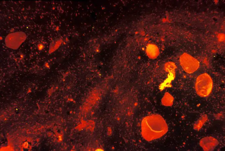
Sputum Microscopy
Culture, Staining, Requirements and Observations
Essentially, sputum is composed of a mixture of saliva and mucus. Also referred to as phlegm, sputum is produced in the bronchi and bronchioles of the respiratory system and coughed up from the lower airways and should not be confused or mistaken for saliva, which is secreted in the mouth for digestion purposes.
It should also not be confused with mucus given that sputum is only produced in parts of the respiratory tract (such as bronchioles). It may vary in terms of color and viscosity ranging from thick to thin or clear to dark yellow in color. All this largely depends on its contents. For instance, blood cells or tar may be present as well as bacteria.
Sputum microscopy is used to determine any underlying health issues for treatment.
Sputum Microscopy
Refers to the microscopic investigation of sputum. This has been shown to be one of the most efficient methods of identifying tuberculosis infection among patients in order to help them start treatment.
For microscopy, sputum may be prepared directly once collected or cultured before microscopy.
Sputum Culture
Culture simply involves encouraging the microorganism in the sputum to grow. The sputum may contain given types of bacteria or fungi.
Using the culture method, these microorganisms are able to grow in a conducive environment, with sufficient nutrition, optimal temperature and moisture etc, making it easier to identify them.
For sputum culture, it is advised not to use mouthwash among other medicinal substances used for mouth washing. This is due to the fact that they may contain various antibacterial substances that would have a negative impact on the final results.
The sample is either obtained by inducing or being coughed up (expectorated) early in the morning before drinking or eating anything.
Depending on the type of microorganism suspected to be present, the following are some of the growth media used in sputum culture:
- Blood agar
- Cysteine Lactose-Electrolyte-Deficient
- Chocolate agar
Selective media for:
- Pseudomonas species
- Fungi and yeasts
- B. cepacia complex
* Once the microorganism starts to grow, it's identified using a microscope.
With regards to microscopy, bright field and fluorescence microscopes can be used for observing and studying the appearance of acid-fact bacilli. As well, gram stain method can be used for the purposes of detecting the presence of fungi.
Bright Field Microscopy
A bright field microscope is the most basic form of microscope illumination techniques with regards to compound microscopes.
For this technique, no special additional accessories are required in a compound microscope, which means that the user will view the specimen as a darker object that is surrounded by a bright field (background).
Ziehl-Neelsen Technique (Acid Fast Bacilli Staining)
Ziehl-Neelsen staining method is an acid fast staining method used to determine the presence of acid fact bacteria such as the Mycobacterium species. The bright field microscopy is used for this technique.
To determine whether a patient has TB, sputum microscopy is used to detect the presence of mycobacterium tuberculosis, which is the bacteria that causes tuberculosis.
Requirements
- Bright field compound microscope
- Staining rack
- Burning stick
- Tweezers
- Slide rack
- Water/distilled water
- 25 percent sulphuric acid
- Carbol Fuchsin
- Methylene blue/Malachite green
- Sample
- Microscope glass slide/cover slip
Procedure
* Make sure to use a pair of gloves when handling biological fluids like sputum.
- Carefully open the container (that contains the sputum sample) and using a laboratory burning stick (dry) obtain and spread a small amount at the central part of a microscope glass slide - Use rotational movement to create a good smear
- Place the slide on a drying rack and allow to dry for about 30 minutes or use a dryer to dry the smear faster
- Pass the slide over the Bunsen burner flame 3 to 4 times to heat fix while avoiding to overheat
- Place the slide on the staining rack and pour the Carbol fuschin stain (to cover the smear) and heat until it starts evaporating - do not overheat
- Allow the slide to stand for between 4 and 7 minutes then wash wish water
- Pour 20 percent sulphulic acid on the smear and allow it to stand for a minute. Repeat this until the smear appears pink in color
- Wash the slide with water and cover the slide with malachite green stain or methylene blue and allow the slide to stand for about 2 minutes
- Wash the slide with water and allow the slide to dry on the drying/draining rack - use a tissue paper to clear the slides and back of the slide
- Observe the slide under the microscope using 100x oil immersion objective
Observation
If the bacteria is present in the sample, students will see pink rods that are either straight or slightly curved while the background will appear bluish in color - the pink rods are the bacteria.
Notes
Acid fast bacteria like mycobacterium have a lipoid capsule that gives their cell walls a waxy appearance. Their cell walls also contain large amounts of mycolic acids and fatty acids. Due to these compounds in their cell wall, acid fast bacteria require a special technique with regards to staining.
During staining, the primary stain is able to penetrate and enter the cell wall of acid fast bacteria because the stain is lipid soluble. Heating also enhances this. When a decolorizer is used (sulphulic acid) acid fast bacteria retain the primary stain while the non-acid fast microorganism loses the stain.
Acid fast bacteria are able to retain the primary stain because of the characteristic of their cell wall. When the counter stain is used, it is readily taken up by the non-acid fast microorganisms, but not the acid fast bacteria.
Fluorescence Microscopy
A fluorescence microscope is a compound microscope that applies the use of fluorescence and phosphorescence to observe the object. With this microscopy technique, the specimen under investigation is the source of light. This technique is also important for sputum microscopy.
Auramine-Rhodamine Method
Also referred to as Truant staining method, Auramine-Rhodamine staining method is one of the methods used for viewing and studying the Acid-fast bacteria (bacilli). This method is not only easy, but also quick to use, which has made it one of the best alternatives to Ziehl-Neelsen technique today.
Requirements
- Auramine Rhodamine Stain
- 0.5% Acid alcohol
- 0.5% Potassium Permanganate
- Microscope glass slide
- Fluorescence microscope (compound microscope)
- Staining rack
- Drying rack
- Water
- Bunsen burner
- Lab burning stick
Procedure
Using a burning stick (or wire loop) obtain and make a small smear at the center of a glass slide
Pass the slide over the Bunsen burner flame 3 to 4 times to heat fix
Flood the slide with Auramine stain and allow to stand for 15 minute - cover the smear with the stain
Rinse the smear with distilled water until no color remains - do not use chlorine water
Flood the slide with Acid alcohol for about 3 minutes
Wash the slide using distilled water
Flood the slide with potassium permanganate for about 2 minutes
Rinse with distilled water and allow to dry
Observe under the microscope starting with low magnification and then high magnification
Observation
If Acid fast bacilli are present in the sample, students will see them appear as yellow or bright orange in color with a dark background.
Notes
Auramine-Rhodamine (primary stain) is a fluorochrome dye that has affinity for acid fast organisms. When it comes in contact with the cell wall of the bacteria, it forms a complex with the mycolic acid present in the cell wall.
Heat fixing enhances this process making it difficult to decolorize when acid alcohol is used. Here, the secondary stain/counter stain used is potassium permanganate.
This stain is used to make other debris non-fluorescent so that they may not be seen when viewing under the microscope. Therefore, only the cells that take up the primary stain can be seen.
Gram Staining
Gram staining method is used for the purposes of distinguishing between gram positive and negative bacteria. This technique has also been use for studying sputum.
Requirements
- Sample (sputum)
- Glass slide
- Bunsen burner
- Compound microscope (bright field)
- Burning stick/cotton swab
Procedure
- Using a burning stick or cotton swab, obtain a small amount of the sample and make a smear at the center of the glass slide - try making a thin slide
- Place the slide on drying rack and allow to dry
- Pass the slide over the flame several times, but avoid overheating. Simply pass it over the flame several times - heat fix
- Flood the smear with crystal violet and allow to stand for about a minute
- Tilt the slide and rinse with distilled water
- Flood the slide with Gram's iodine for about one minute
- Tilt slide and rinse with water
- Tilt the slide and apply the alcohol drop by drop until it runs clear (95 percent ethyl alcohol/acetone)
- Rinse with water
- Flood the slide with safranin for about a minute
- Tilt slide and rinse with distilled water
- Blot the slide dry
- View under the microscope under high power (oil-immersion)
Observation
When viewed under the microscope, students may observe different types of cells (Squamous epithelial, mononuclear cells, respiratory epithelial cells etc) and other cells like Streptococcus pneumoniae, staphylococci and Hemophilus influenzae if they are present.
While the body cells may appear pinkish, the microorganisms will appear darker having retained the primary color due to their thick peptidogycan layer on their cell wall.
* By observing and studying the types of cells and microorganisms present in the smear using gram-staining, it's possible to tell the patient's type of infection.
Related: Blood Smear, Urinalysis, Microscopy Culture and Sensitivity
Return to Microscopy Applications
Return from Sputum Microscopy to MicroscopeMaster Research Home
References
Dr. Annette Jepson (2008) Laboratory Techniques for the Diagnosis of TB – what’s available?
D. J. Flournoy (1999) Interpreting the Sputum Gram Stain Report.
http://www.sciencedirect.com/topics/medicine-and-dentistry/acid-fast
http://www.stoptb.org/wg/gli/assets/documents/TB%20MICROSCOPY%20HANDBOOK_FINAL.pdf
Find out how to advertise on MicroscopeMaster!

![Haemophilus influenza Gram Stain by Bobjgalindo (Own work) CC BY-SA 4.0-3.0-2.5-2.0-1.0 (https://creativecommons.org/licenses/by-sa/4.0-3.0-2.5-2.0-1.0)], via Wikimedia Commons Haemophilus influenza Gram Stain by Bobjgalindo (Own work) CC BY-SA 4.0-3.0-2.5-2.0-1.0 (https://creativecommons.org/licenses/by-sa/4.0-3.0-2.5-2.0-1.0)], via Wikimedia Commons](https://www.microscopemaster.com/images/512px-Haemophilus_influenzae_Gram.jpg)


