Sperm Cells
** Definition, Function, Structure, Adaptations & Microscopy
Definition: What are Sperm Cells?
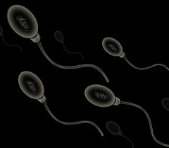
Sperm cells are gametes (sex cells) that are produced in the testicular organ (gonad) of male human beings and animals.
Like the female gamete (oocyte), sperm cells carry a total of 23 chromosomes that are a result of a process known as meiosis. In both animals and human beings, among many other organisms, these cells are involved in the sexual mode of reproduction which involves the interaction of male and female gametes.
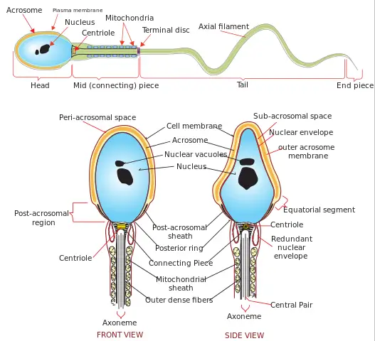
The general morphology of sperm cells consists of the following parts:
- Distinctive head
- Midpiece (body)
- Tail
Structure and Function
Before looking at the structure and function of sperm cells, it's important to understand the process involved in their production (spermatogenesis).
Spermatogenesis
In male animals, the hypothalamus plays a crucial role of monitoring the level of testosterone in blood. Low level of the hormone indicates low testicular activity, which triggers the hypothalamus to release releasing hormone known as gonadotropin-releasing hormone (GnRH).
GnRH then flows to the pituitary gland and stimulates the production of luteinizing hormone (LH) and the follicle stimulating hormone (FSH).
From the pituitary gland, the luteinizing hormone surges and stimulates leydig cells present in testicles to produce testosterone. Follicle stimulating hormone, on the other hand, plays an important role of concentrating this hormone in the seminiferous tubule to begin sperm formation.
In the inner walls of the seminiferous tubules, a group of cells known as spermatogonial germ go through a mitotic division to produce primary spermatocytes (haploid). These cells then undergo meiotic division resulting in the production of secondary spermatocytes. The spermatocytes then undergo second meiotic division to form spermatids that develop to form mature sperm cells.
* Spermatogenesis takes about 74 days to complete
* There are two main processes involved in spermatogenesis. The first process (meiosis) reduces chromosomes to half while the second involves changes in size and shape as the sperm mature to their normal shape.
Structure
While their general morphology includes a head, body, and tail, all sperm cells do not necessarily look alike.
As a result of various abnormalities, they may vary in shape and size while other differences may be observed on any part of the cell (head, body, tail).
A normal sperm will have the following characteristics:
- A smooth oval head - The head of a normally formed sperm has a smooth surface and resembles the shape of an egg
- The head of the sperm measures 2.5 to 3.5 um in diameter and 4.0 to 5.5 um in length (um=micrometers). This results in a 1.50 to 1.70 length to width ratio
- They have a well-developed acrosome that covers 40 to 70 percent of the oval shaped head
- A slim middle section (body) that is approximately the same length as the head
- A thinner tail section that is about 45 micrometer in length
A sperm cell consists of a head, body (mid-section) and a tail. Each of these parts is equipped with various molecules and smaller structure that allow the sperm as a whole to function properly.
Sperm Head
As already mentioned, a normal sperm head has a smooth and oval shape. The head section also resembles an egg due to its broad base and tapering apex.
The head is the most important part of the cell given that it contains the nucleus (genetic material with 23 chromosomes) required to form a new organism.
Apart from the nucleus, the head is also made up of a several parts that include:
Acrosome and Acrosomal Cap
Together, the two (acrosome and acrosomal cap) make up the acrosomal region. Formed during spermiogenesis, the acrosome is the product of Golgi complex and contains a number of contents such as acrosin enzyme in the acrosomal matrix. Apart from the enzymes, the acrosome also contains such polysaccharides as mannose, hexosmine and galactose.
The acrosome occupies the space between the interior plasma membrane and nuclear membrane. The acrosome itself has an inner and outer membrane (acrosomal membrane) where the outer membrane borders the plasma membrane while the inner acrosomal membrane borders the nuclear membrane.
The acrosome plays a number of important roles in fertilization. For instance, with a number of its associated molecules, the acrosome is involved in the recognition of the oocyte (egg) to be fertilized.
Once the sperm cell comes in contact with the diffusible molecules from the egg jelly, this stimulates the cell to swim towards the eggs. This recognition of the egg based on molecule composition is known as chemotaxis.
Having identified a high concentration of the molecule, the cell swims towards the egg (area of high molecule concentration) and makes physical contact. In turn, physical contact results in acrosome reaction.
* Chemotaxis allows the sperm to navigate towards the eggs through chemical signals. Therefore, this is an important process that ensures that the sperm fertilizes a conspecific egg (within the same species).
* Primary ligands (proteins) located near the acrosome recognize the target gamete.
Acrosome Reaction
The acrosome reaction is an important event that occurs when the sperm comes in contact with oocyte membrane at different sites.
For instance, in some animals, sperm contact with zona pellucida on the plasma membrane of the oocyte initiates acrosome reaction. This is a calcium-dependent event that results in exocytosis (action in which cell molecules are released from the cell) of the outer acrosome membrane thus exposing contents (enzymes) of the acrosome.
This allows acrosome enzymes (e.g. acrosin) to be released and support sperm entry into the egg. Acrosin/proacrosin, one of the secondary ligands, is involved in lysis of the thick membrane covering the ovum (zona pellucida)
Essentially, the enzyme (acrosin) is stored in the acrosome in an inactive form known as zymogen. The pH level inside the acrosome is lower which causes the enzyme to remain inactive.
When it comes in contact with the glycoproteins of the ovum membrane (zona pellucida), the enzyme is converted into acrosin, an active form that is capable of acting on the membrane. This, in turn, allows the sperm cell to penetrate and enter the egg for fertilization to take place.
* Acrosome enzymes are also known as lysosomal enzymes.
Nucleus - The sperm head is the part of the cell that contains the nucleus. The nucleus takes up 65 percent of the head and consists of 23 chromosomes.
Once the sperm cell enters the egg, the chromosomes combine with the female gamete to make up 46 chromosomes - It's the total of 46 chromosomes that determine the characteristics of the new organism (fetus etc).
* The sperm head makes up about 10 percent of the entire cell.
Midpiece
The midpiece is the central part of the sperm cell between the head and the tail. Like the head, the midpiece makes up about 10 percent of the total sperm length. Unlike the sperm head that carries genetic material, the midpiece contains tightly packed mitochondria that provide the energy requires for swimming.
In addition to providing the energy required for swimming, mitochondria is also suggested to play a role in controlled cell death known as apoptosis.
Centriole - The centriole is part of the sperm cell located between the head and the midpiece. In a complex referred to as the centriole-centrosome complex, the centriole is involved in the formation of sperm aster and zygote aster.
These are essential for movement of the pronuclear for union with the female genome. Moreover, the centriole is involved in the production of mitotic apparatus involved in separating chromosomes during cell division while at the same time being the template for all subsequent centrioles.
Tail
The sperm tail is a thin, elongated structure that makes up about 80 percent of the entire length of the sperm.
While the tail may appear to be one long continuous structure, it is divided into several parts that include:
- Connecting piece – This is the part that connects the flagellum to the sperm head
- Midpiece - In some books, the midpiece is described as part of the tail. It contains mitochondria and thus provided the energy required for movement
- Principal piece (axial filament)
- End piece
* The principal piece and the end piece of the flagella help generate the waveform that allows for movement.
Motility
Motility is one of the main characteristics of a well developed sperm cell. In mammals, two types of physiological motility have been identified.
These include:
Activated motility - This is the type observed in the early stages of motility (in the epididymis as well as freshly ejaculated sperm). In this type of motility, the sperm's flagella beats gently from one side to another as the cell moves along what may appear to be a straight path.
Hyperactivated motility (hyperactivation) - Hyperactivated motility is the second type of physiological motility. Compared to activated motility, this type of motility occurs is in the female reproductive tract (site of fertilization).
Hyperactivated motility is also more erratic, with the flagellum depicting a symmetrical, lower-amplitude waveform. Due to the erratic pattern of motion in hyperactivated motility, more energy is used for movement.
* Hyperactivated motility serves to prevent the sperm cell from getting trapped, propelling through the reproductive tract (of the female) as well as enhancing sperm penetration into the egg (oocyte).
* Motility is only possible if the flagellum is well developed and fully functional and if the cell has a source of energy to support movement.
* Sperm cells have been shown to swim at an average rate of 3mm a minute.
Axoneme and Molecular Mechanism of Motility
The axoneme is the central strand of the tail (flagellum). It's one of the main structures of the flagellum and is commonly known as the motility motor. The axoneme is made up of structures referred to as microtubule doublets (containing inner and outer axonemal dynein) and a central pair (9+2 structure) and extends from the connecting piece of the tail to the end piece.
Within the flagellum, the microtubules (nine microtubule doublets) are connected by nexin links. In addition, they are linked to the central pair through radial spokes. These projections (radial spokes) also play an important role of aligning the microtubules around the central pair.
During motion, dynein in the microtubules causes the microtubule to slide in relation to the adjacent microtubules, which promote motility. With the energy provided from the mitochondria (ATP energy), axonemal moves towards the flagellum base, which causes the microtubule to slide down.
Given that the microtubules are connected to the connecting piece located behind the head, there is some resistance to the movement which in turn causes the flagellum to bend. Through this action, the flagellum forms a whip like bend.
Movement, however, is promoted by several other actions that include:
- Detachment of the dynein from an adjacent microtubule
- Processes take place at one side of the axoneme
Adaptations of Sperm Cells
- Streamlined body - The sperm has a streamlined body that allows it to move rapidly to reach the target egg cell. For instance, the head has a tapering apex which helps reduce drag as the cell travels in the female reproductive tract.
- Tightly packed mitochondria - The midpiece of a sperm carries about 70 mitochondria, which is the source of energy (ATP). This provides sufficient energy required for propulsion as the cell travels towards the female gamete. The mitochondria of sperm cells is discarded once the sperm head penetrates the egg.
- Basic amines - The sperm contains a number of basic amines such as cadaverine and spermine among others. These amines are responsible for the alkaline (slightly basic) nature of semen that protects the sperm. Given that the vaginal canal is acidic, the amines protect the DNA from denaturation thus promoting successful fertilization. See more on amines.
- Acrosome - As already mentioned, the acrosome plays an important role in chemotaxis to identify target female gamete and contains lysosomal enzymes that degrade the thick membrane of the egg. The acrosome therefore helps promote fertilization.
Spiral Nanostructure
Recently, researchers from the University of Gothenburg discovered a spiral nanostructure located inside the microtubules at the tip of the flagellum.
Measuring about a tenth of the tail, the structure is suggested to serve as a cork within the microtubules that stops them from growing and shrinking.
Microscopy
Microscopy is one of the methods used in analysis. Using a simple wet mount procedure, it is possible to observe the morphology, population as well as the movement of sperm cells under the microscope.
Requirements
- A compound microscope (phase contrast microscope or differential interference contrast microscope)
- Sperm sample
- Warm extender or buffered saline
- Microscope slide (pre-warmed)
- Coverslip
Procedure
- Dilute sample in warm buffered saline or extender
- Using a pipette, place about 20 ul of the mixture on the microscope glass slide (a pre-warmed glass provide favorable conditions for the sample)
- Using a cover slide, gently cover the sample by lowering the cover slip at an angle in order to remove air bubbles
- Mount and view the slide under the microscope starting with low power
* Using this technique, it is possible to view the general morphology of sperm cells as well as sperm motility.
* This particular technique is recommended to observe live sperm cells and sperm motility.
* Phase contrast or differential interference contrast microscopes offer good contrast that makes it possible to recognize the sperm cells under the microscope.
Staining
Compared to a wet mount (that is less likely to cause damage to the cells) staining allows for better differentiation thus making it possible to view different regions of the sperm cell. The problem with this, however, is that it kills sperm cells.
Procedure
- Using a cotton swab, prepare a diluted smear on to a clear glass slide
- Dip the slide into a fixative for about 5 minutes to fix the smear
- Using a heating plate, dry the smear for about 15 minutes at about 37 degrees Celsius
- Dip the slide in tap water and then dip in stain A (Spermac A) for about a minute
- Dip the slide in water and then dip the slide in stain B (Spermac B) for about one minute
- Dip the slide in water and then dip in stain C (Spermac C) for about one minute
- Wash the slide by dipping in tap water and allow the slide to dry for about 12 hours
- Mount the slide and view under oil immersion under high power
Observation
Staining makes it possible to clearly recognize all parts of the sperm cell. Here, it is also possible to identify any defects of the sperm
* Sperm cells appear red in color while the acrosome, centerpiece and the tail appear green.
Take a look at Sertoli Cells as well as Leydig Cells
Return from Sperm Cells to MicroscopeMaster Home
References
Christopher J. De Jonge and Christopher L. R. Barratt. (2017). The Sperm Cell: Production, Maturation, Fertilization, Regeneration.
Damayanthi Durairajanayagam et al.. (2015). Sperm Biology from Production to Ejaculation. ResearchGate.
Ryuzo Yanagimachi. (2011). Mammalian Sperm Acrosome Reaction: Where Does It Begin Before Fertilization? Oxford Academic.
Links
https://onlinelibrary.wiley.com/doi/abs/10.1002/9781118904398.ch118
Find out how to advertise on MicroscopeMaster!


