Phylum Protozoa
** Classification, Structure, Life Cycle and Microscopy
Introduction
Essentially, protozoa are single-celled eukaryotes. This means that they are single celled organisms that have a nuclei as well as a number of other important organelles within the cytoplasm and enclosed by a membrane.
They exist as free-living organisms or as parasites. This makes this phylum a diverse group of unicellular organisms, varying in shape and size.
Examples include:
Anatomy (Bodily Structure)
Given that they are eukaryotes, protozoa are larger cells of between 10 and 100 micrometer in diameter (compared to prokaryotes) with a more complex structure. This means that they have a cell membrane which bounds the organelles, a DNA that is also bound by a membrane, nucleoli, ribosome, Golgi apparatus and multiple linear chromosomes with histones among others.
It's worth noting that organelles present in these cells will vary from one type to another.
There are also a number of organelles that are exclusive to protozoa, these include:
- Trichocysts of Paramecium
- Certain skeletal structures
- Contractile vacuoles
Compared to other ciliates, the nucleus of protozoa is vesicular. As such, the chromatic is scattered resulting in a nucleus that is diffuse in appearance. However, this also varies from one to another. For instance, in the Phylum Apicomplexa, the vesicular nucleus had one or more nucleoli with DNA while the endosone of trypanosomes are lacking DNA.
Protozoa also have in place locomotory structures such as pseudopodia, flagella and cilia which are used for movement. These structures are also surrounded by the plasma membrane.
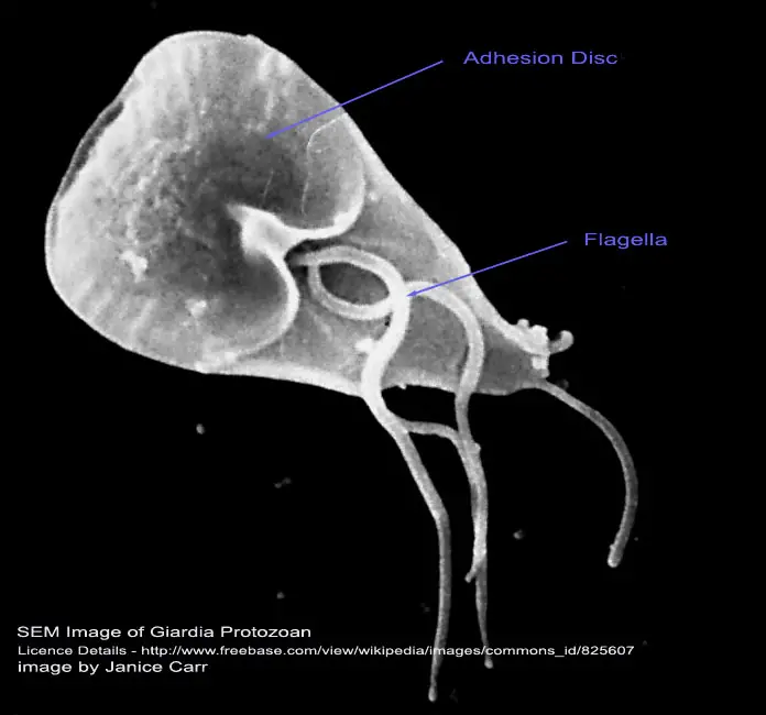
As well, the pellicle (outer surface of some like the Giardia) is rigid enough to support and maintain a distinctive shape while at the same time allowing for twisting and bending when moving.
Classification of Phylum Protozoa
Because of their diversity, protozoa present several problems when it comes to classification. They are considered to be under the sub-kingdom protista with more than 50,000 species being described as free-living (these are the type that do not directly depend on others for survival).
Free-living protozoa can be found in virtually every possible habitat. Based on both light and electron microscopy morphology, they have been classified into six major phyla with a majority of disease causing protozoa falling under the phyla Sacromastigophora and Apicomplexa.
The following are some of the sub-phyla and classes within these sub-phyla based on locomotive structures:
Plasmodroma - The locomotive structures of this sub-phylum may be flagella, pseudopodia or none at all. Classes that fall under this sub-phyla include; Mastigophora (use one or more flagella for locomotion), Sarcodina (used pseudopodia for locomotion and for capturing food) and Sporozoa which lack locomotive structures.
Ciliophora - These are in the Sub-phyla Ciliophora use cilia or sucking tentacles in some stages or throughout their life span. Ciliata (which use cilia throughout) and Suctoria (which use cilia when young and tentacles as adults) are some of the class that fall under this sub-phylum.
Sarcomastigopohora - The locomotive structures used in this sub-phylum include pseudopodia or flagella. Here, the nuclei is also of one kind (monomorphic). Super class Mastigophora, which falls under this sub-phyla are flagellates and thus use flagella for locomotion.
The Phytomastogophoerea also falls under this sub-phyla and use flagella in some cases. Under the Class Phytomastogophoerea is Order Chrysomonadida, which includes such organisms like Chrys amoeba, synura and ochromonas among others.
** These are just a few in the classification. It is extensive and contains many more organisms.
Classification Based on Mode of Existence
Of the existing protozoa, there are about 21,000 species that occur as free-living in a variety of habitats while another 11,000 species occur as parasitic microbes in both vertebrate and invertebrates hosts.
The free-living species can be found in various habitats and particularly in soil and water. These types of protozoa have little impact on human health given that they do not directly depend other organisms for their survival. However, some of the free-living can cause pathology when introduced into a human host.
Others will also affect human health by producing toxins.
The following are some of the free-living amoebae that can also cause human disease:
- Naegleria fowleri - This species is mostly found in moist soil and can be located all across the world. It causes acute primary amebic meningoencephalitis.
- Acanthamoeba - Found in soil and water, acanthamoeba can cause chronic granulomatous amebic encephalitis, amebic keratitis, granulomatous skin as well as lung lesions.
- Balamuthia mandrillaris - Causes sub-acute to chronic granulomatous amebic encephalitis as well as granulomatous skin and lung lesions.
- Sappinia diploidea
Parasitic Protozoa
Parasitic protozoa are the type that depend on the host for survival. As such, they live inside the host and even cause health problems.
The following are some of the parasitic:
Sarcodina ( e.g. Entamoeba) - Entamoeba histolytica is a type of amoeba that lives in the human alimentary canal. For most part, they are harmless and feed on various bacteria and particles that may be present in the intestine.
Although they are mostly harmless, this parasite may invade the intestinal wall or the rectum where they cause ulcerations and even bleeding along with pain, vomiting, and diarrhea among other symptoms.
Trypanosomes - This is a flagellate that lives in the blood stream. Various species of this parasite cause such diseases as:
- sleeping sickness,
- leishmaniasis
- Chaga's disease
Mastigophora (e.g. Giardia) - This is a flagellate that is mostly found in the small intestine of the host. The giardia typically attach themselves on to the intestinal lining causing inflammation, diarrhea as well as abdominal pain among other types of symptoms.
Sporozoa (e.g. Plasmodium) - The plasmodium species is a parasite that lives in the blood stream of human beings, Once in the red blood cells, the parasite feeds on their cytoplasm. As they continue multiplying within the cells, this causes the cells to burst which in turn results in many more parasites being released into the circulatory system.
Life Cycle
Parasitic Protozoa
For the parasitic forms, the life cycle stages may occur intercellular, intracellular or in the lumen of given organs. Because of the diversity, it is not possible to describe a single or one common life cycle sequence. Here, therefore, we shall look at three of the most common broad patterns exhibited by this group of protozoa.
First pattern:
This pattern is common in the phylum Apicomplexa and involves an alteration between asexual and sexual reproductive stages.
The process starts with the cycles of asexual reproduction where the cycles of schizogony (involving mitosis and cytokinesis) in the tissues of the host results in increase population.
Following this stage, some in the population start undergoing gametogony (a sexual process) to produce gametes. These gametes then unite and divide asexually to produce sporozoites through a process known as sporogeny.
It is these sporozoites that are then capable of infecting a new host and the process continues. Here, it is worth noting that transition in to a new host is through cysts, which are tough under stressful conditions. The cysts can survive external conditions (outside the body) and contain the sporozoites.
Once in a new host, the sporozoites start the reproduction cycle again. Some of the species in this phylum (Apicomplexa) require two hosts to complete their life cycle. This includes a vertebrate host where the parasite goes through schizogony and gametogony and an invertebrate where the gametes unite and sporiogony occurs in the tissues.
Second pattern:
The second pattern is common among most flagellates and involves asexual reproduction. For these, a number of morphological transformations occur during the cycle. However, they all reproduce through binary fission.
Some of the species in this group will complete this cycle in a vertebrate host as they transmit from one host to another through cysts, which can survive tough conditions better. Therefore, as in the case with the Apicomplexa phylum, some species in this group will also require two hosts to complete their life cycle.
Third pattern:
This is particularly common among amoebas and involves asexual reproduction. Unlike the others, a single host is required to complete the reproduction cycle. Here, trophozoites in the host live in the lumen of the gut and continue to multiply through binary fission.
Here, under certain conditions, the trophozoites may be stimulated to encyst as they undergo nuclear division within the cyst. Once the cyst is ingested by another host, the cycle continues.
Life Cycle of Free Living Protozoa
For this group, the life cycle largely involves the growth and increase in size of the organism which is then followed by binary fission (or other forms of asexual reproduction).
For the free-living, sexual reproduction only occurs under unfavorable conditions (unfavorable temperature, or reduced food supplies etc). However, these factors often vary from one species to another.
During the growth and division cycle of the free-living protozoa, there is a phase of DNA synthesis, chromosome replication as well as the growth of the cells.
The phases of the cycle include:
- First division phase
- End of division phase and beginning of DNA synthesis
- DNA synthesis
- End of DNA synthesis and beginning of next division
Classification based on Nutrition (How they Obtain Energy)
There are three main categories based on nutrition.
These include:
- Photo-autotrophs
- Photo-heterotrophs
- Chemoheterotrophs
Autotrophs like some of the flagellates synthesize carbohydrates from carbon dioxide and water using chlorophyll. Here, radiant energy from the sun is used.
Most of the photoautotrophic flagellates including members of Euglenida, Cryptomonadida as well as Volvocida also tend to combine autotrophy with heterotrophy. For this reason, they are often described as acetate flagellates.
Some of their source of carbon include acetates, simple fatty acids as well as alcohols. While they are autotrophs in the light, these flagellates switch to heterotrophs in the dark.
A majority of the free-living protozoa fall under this category. As such, they depend on a wide range of diet. Whereas some feed on bacteria (microbivores) others feed on algae and are described as herbivores. The carnivorous feed on both of the two trophics (herbivores and microbivores).
The free living are also divided into two groups (morphological). These include those with a mouth/cytostome and those that lack a mouth or a definite point of entry for food. For instance, whereas some flagellates and many ciliates (apart from some apostomatida) have a cytostome the Sarcodina lack a mouth.
Chemoheterotrophic - This group includes those that require energy and organic carbon sources.
Microscopy
As previously mentioned, protozoa are very diverse. As such, they are distinguished from one another based on their different structural features, means of locomotion as well as the formation of spores.
Using a light microscope, it's possible to view different types of protozoa.
Sample collection
Protozoa can be obtained from almost any given habitat. Whereas the free-living species can be found in water as well as various moist habitats, the parasitic can be found in most metazoan (developed animals).
For students, it would prove easier to use the free-living protozoa, which can be obtained from such habitats as mud, ponds and transient bodies. Here, it is worth noting these are very fragile. For this reason, they should be handled with care.
It is also important to be careful given that even free-living protozoa can become parasitic. Protozoa can also be cultured in order to increase their numbers for observation. Some of the medium used include split pea (for Eglena) distilled water with wheat grains (for chilomonas) as well as hay (for peranema) among others.
Microscopic Observation
Some of the requirements for microscopy include:
- A microscope
- Microscope slides
- Microscope clips
- Distilled water (or tap water)
- Dropper
Wet Mounting Technique
Wet mounting technique is the technique that simply introducing the sample/specimen on to a drop of water and viewing it under the microscope.
If the sample was obtained from a pond, then the following process can be used:
- Gently shake the container (to distribute the protozoa in the water)
- Using a dropper, obtain a sample of the pond water from the container
- Place a drop of the sample onto the center of a microscope slide and cover with a cover slip (always ensure that the microscope slide and slip are clean to avoid introducing other microorganisms)
- Place the slide onto the microscope stage for viewing
In some cases, staining may be used to increase contrast and get a clearer view. Some of the stains used here include:
- Bismarck Brown
- Brilliant Cresyl Blue
- Bromothymol Blue
- Carmine Powder
- Methylene Blue
More on Cells:
Eukaryotes - Cell Structure and Differences
Prokaryotes - Cell Structure and Differences
Diatoms - Classification and Characteristics
Protists - Discovering the Kingdon Protista in Microscopy
Fungi - Mold Under the Microscope
What does Phylum mean in Biology?
Specifically learning about Vorticella, Rhizopoda
Take a more in-depth look at Parasitology as well
See Amoeba under the Microscope specifically Acanthamoeba
Read about Parasites under the Microscope here
Also check out Microrganisms, especially in Pond Water.
Take a look at Microscope Slide Preparation.
And if in need of a microscope then be sure to read our Darkfield Microscope Buyer's Guide and Phase Contrast Microscope Buyer's Guide.
Look at Protozoology as field of study
Return to Cell Biology - Components, Cycles, Processes and Microscopy Techniques
Return from Protozoa to Best Microscope Information and Research
References
Ward's Science (2005) Working with protozoa.
Johanna Laybourn-Parry (1984) A functional biology of free-living protozoa.
Gary N. Calkins (1906) The Protozoan Life Cycle.
J. P. Kreier and J. R. Baker (1987) Anatomy and physiology of the protozoa.
R. W. Hegner (1926) Homologies and Analogies Between Free-Living and Parasitic Protozoa.
Martinez AJ, Visvesvara GS (1997) Free-living, amphizoic and opportunisitic amebas. Brain Path. 7:583-598.
Mackean & Ian Mackean (2017)Parasitic Protozoa, an Introduction.
Links
http://parasite.org.au/para-site/contents/protozoa-intoduction.html
Find out how to advertise on MicroscopeMaster!
![By Donald Hobern from Copenhagen, Denmark (Protozoa sp.) [CC BY 2.0 (http://creativecommons.org/licenses/by/2.0)], via Wikimedia Commons By Donald Hobern from Copenhagen, Denmark (Protozoa sp.) [CC BY 2.0 (http://creativecommons.org/licenses/by/2.0)], via Wikimedia Commons](https://www.microscopemaster.com/images/Protozoa1stpic.jpg)


