- Phytopathology -
Analysis Using Fluorescent and Electron Microscopy
Phytopathology is the study of the causes of disease, whether environmental or biological, among plants. This field of science determines what pathogen is present and then investigates where it came from, how it affects a plant, and why it only harms some plant species.
Upon obtaining this knowledge, scientists can then work to create prevention and control methods so that the disease does not spread or continue to damage the already infected host.
Healthy versus Unhealthy
Like human beings, plants carry out specific life functions when they are healthy but stop performing some tasks or engage in new ones when an infection is present.
A healthy plant can carry on cell division, has the ability to take in nutrients and water and transport them to all parts of the organism. It then breaks down and stores the nutrients so the cells can continue their essential functions, including the production of seeds and maintenance of reproductive organs.
If a plant cannot perform any one of these functions, it has a disease or is unable to survive in its current environment.
Since pathogens start in a single cell or tissue space, it is impossible to notice the onset of the disease, which allows it to spread throughout the organism, eventually causing external changes.
Phytopathology taught scientists that the yellowing of leaves, known as chlorosis, wilting, stunted growth or blight, which is a quick death of the plant or its parts, are all some of the symptoms a diseased plant might display.
Additionally, an observer might also notice scorching at the attachment site of the leaf, detachment of seedlings, a condition known as damping off, or necrosis, which is the complete death of plant tissues and cells.
Plant Pathogens and Disease
Discoveries made through phytopathology include numerous plant diseases and the pathogens that cause them, whether biological or environmental. Some of the environmental factors that lead to diseased plants involve light, minerals and nutrients, temperature, moisture, pesticides and pollution, oxygen and soil.
If a plant receives too much or too little of certain external elements, experiences a rapid or harsh change in its surroundings or has exposure to poisons, it becomes sick.
Biologically, plants are just as susceptible to bacteria and viruses as people, with the only difference being the type of pathogen that causes illness.
In plants, parasites, protozoa, fungi, viroids, prokaryotes and nematodes are all responsible for disease.
See also bacteria like Pseudomonas and Xylella Fastidiosa.
Phytopathology shows that any of these factors may lead to problems such as tumors, which is when a cell becomes abnormally large; hyperplasia, a condition that causes rapid cell division of the infected cell; or for an organ to become irregular, as in crown gall disease.
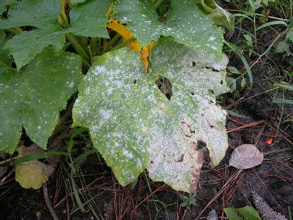
Powdery mildew, a Biotrophic Fungus
From Bioencyclopedia.com
Microscopic Analysis
When engaging in phytopathology, one of the most important tools to use is the microscope, which led to numerous discoveries throughout history, despite being previously underused in this field.
The basic microscope is sufficient for viewing larger material like cells, tissues, and organelles but it is not strong enough to see the smaller material hidden within these structures.
Fortunately, technology fixed this problem, as new, improved microscopes allow scientists to view material previously undiscovered, analyze it and document their findings, which could make a significant contribution to medicine, farming or pharmacology.
Fluorescent Light Microscopy
The major difference between a light microscope and a fluorescent one is the intensity of the light used, which provides a very different and highly detailed result.
As the name suggests, the fluorescent microscope works by using a fluorescent chemical, species or specific part of an organism that is naturally fluorescent, such as chlorophyll, making it a beneficial tool in phytopathology.
The fluorescent light microscope has the ability to manipulate visible and fluorescent light, filter radiation based on wavelength, and then separate the low and bright light to obtain an image.
This process occurs by radiating the test sample with excitation light that has the same wavelength as the specimen, thus causing it to gain energy.
Once the sample returns to its normal energy level, it emits a weak light that requires separation from the bright excitation light so scientists can study the image created by the fluorescing areas.
The use of this tool in phytopathology is very important, as it contributed to the study of various types of plant cells and the enzymes, along with their pathway, that destroy cell walls during an infection.
It also assisted in the discovery of a pattern in some plants that marks the location of germ tube emergence and suggests that proteins at these sites affect the ability of an infectious cell to interact with a healthy plant surface at the biological level.
Electron Microscopy
In addition to the fluorescent light microscope, the electron microscope also plays an important role in phytopathology, as it allows researchers to observe other materials within an organism, tissue or cell at a micro-level.
The electron microscope is higher in magnification than a traditional light microscope, as it can increase the image of an object by two million times its normal size.
The basis for its design is the correlation between resolution and wavelength, which also incorporates the use of electrons to create the final image.
An improvement to this type of microscope is the scanning electron microscope, a device that allows the observer to view their sample as a three-dimensional object.
This technology gave researchers the opportunity to observe a fungus penetrate a plant cell, grow, colonize, and reproduce within a couple of days. It also showed the pathway of the fungus and the internal destruction it caused to the plant.
The scanning electron microscope also enabled scientists to study a mycoparasite and the hyphae of a fungus.
The findings showed a significant difference in size, attachment method to the host cell and process of infection, as well as the attachment site, destruction at the site and detachment of the infectious cell from the host.
Phytopathology is an important scientific field that is not yet highly credited, mostly because so scientists know so little about plants, their diseases and how they affect, or could possibly benefit, humanity.
However, the technological tools now available are allowing research to take on a new direction and the amount of new knowledge is quickly growing.
Once researchers have a better understanding of plants and their pathogens, they can begin to apply or compare the information to human studies and eventually find solutions to various health problems plaguing both species.
Other Pathology Articles from MicroscopeMaster:
See Differences between Plant Cells and Animal Cells
Return from Phytopathology to Microscopy Applications
Return to Microscopy Research Home
Find out how to advertise on MicroscopeMaster!


