Phylum Basidiomycota
** Characteristics, Reproduction and Life Cycle
Overview
The phylum Basidiomycota is a large group that consists of over 30,000 species. These include puffballs, jelly fungi, toadstools, and stinkhorns, etc. which can be found in terrestrial and some aquatic habitats.
The majority of these species are free-living saprophytes but some exist as parasites of trees and some plants while others form symbiotic relationships with their hosts. While they can cause significant damage, basidiomycetes also play an important role in the recycling of nutrients in nature.
Currently, the group is divided into three major subdivisions (subphyla) that include:
- Agaricomycotina - Consists of over 21,000 species
- Pucciniomycotina - Consists of 8,400 species
- Ustilaginomycotina - Consists of 1,700 species
Phylum Basidiomycota Characteristics
Agaricomycotina
Agaricomycotina is a large phylum that makes up about 70% of all phylum Basidiomycota. It's also estimated to make up about a third of all fungi.
Some examples of species in this group in phylum Basidiomycota include:
- Halocyphina villosa
- N. globospora
- Mycaureola dilseae
- Nia vibrissa
- Digitatispora lignicola
- Gloiocephala aquatica
* The subphylum Agaricomycotina was formerly known as Hymenomycetes.
Morphology
Due to its large size, Agaricomycotina is a diverse group that comprises jelly fungi, mushrooms, and yeasts. For this reason, members of the group exhibit significant variation in structure and general morphology.
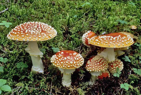
Of over 21,000 Agaricomycotina species, about 16,000 of those form mushrooms. They are characterized by a stalked-umbrella morphology with gill-like structures (lamella) from which spores are produced.
Some of the other parts of a mushroom include the volva, cap/pileus (umbrella-shaped), tubes, and mycelia threads.
Compared to mushrooms, jelly fungi (e.g. Auricularia cornea and Tremella auricularia) do not have a stalk/stem and have a gelatin-like consistency. They also lack gills found in mushrooms and display a variety of colors from pink and orange to brown, black, and white. Yeasts, on the other hand, are unicellular organisms that can be seen with the naked eye in colonies.
* All Agaricomycetes are characterized by a dolipore. This is a septum with a barrel-shaped swelling around the pore. While septal pores can be found in Pucciniomycotina and Ustilaginomycotina species, they do not have a septal core cap.
Apart from the three mentioned morphologies, the group also consists of puffball fungi (e.g. Aseroe rubra). These are classified in the class Agaricomycetes and are characterized by a ball-like/spherical shape when they mature.
Like jelly fungi, puffballs do not have a cap (umbrella-like structure) and gills. However, they still produce spores in their life cycle. Here, spores are produced at the part known as the gleba. Depending on age and species, they vary in size and color (yellow, pinkish, white, cream).
* Some mushrooms can grow to be over 40 cm in diameter.
Habitat/ecology - Agaricomycotina species can be found in various terrestrial and aquatic habitats. Agaricomycetes, which make up over 80 percent of all Agaricomycotina, can be found in a variety of terrestrial ecosystems where they cause wood decay.
Given that most species cause wood decay, they can be found in large numbers in forest areas/forest covers. A few species are aquatic where they also exist as decayers.
* A few Agaricomycetes are pathogens and others form a mutualistic relationship with some plants.
Dacrymycetes are also commonly found on land where they form associations with different types of plants. However, members of this group also cause decay.
Tremellomycetes are also land-based species found in association with plants. However, yeast species can be found in some aquatic environments, glacial mountains, as well as polar habitats.
Reproduction and Life Cycle of Agaricomycotina
Mushrooms (Coprinus comatus)
Also known as the Shaggy Mane Mushroom, Coprinus comatus is one of the most studied members of the subphylum Agaricomycotina.
Spore - The life cycle of the mushroom begins with a spore (basidiospore). Basidiospores vary in shape depending on the species (subglobose to oval) and range between 6 and 8 um in length and 5 to 7 um wide. They also have a thin outer wall and a single nucleus. These spores are released into the environment and germinate when they fall on the right substrate under the right conditions.
Hyphae - Once the spores land on the proper substrate under the right conditions (moisture and oxygen at about 30 °C), it germinates giving rise to structures known as hyphae. As the hyphae elongate and branch, the nucleus within each cell replicates into two and is divided by a separating wall (dolipore septum).
As mentioned, these septa are characterized by central barrel-shaped canals (these canals are located at the center of each septum). Located along the length of the hyphae, these septa have two main functions namely, to allow for movement of cytoplasm along the hyphae and prevent the nuclei from moving between compartments.
Homokaryotic mycelium - As the nucleus continues to divide and the hyphae continue elongating and branching, they give rise to a hyphal network known as a homokaryotic mycelium.
Basidiocarp - For the most part, basidiocarps are only produced when homokaryotic mycelium fuse with genetically compatible mycelium. Here, the hyphae from each of the mycelia fuse through a process known as plasmogamy.
In doing so, they produce a hyphal compartment that contains pairs of unique nuclei (with nuclei from the respective parent mycelia).
Secondary hyphae/heterokaryotic mycelium - Within the hyphal compartment, one of the nucleus (from the pair) divides and gives rise to two daughter nuclei. This is followed by the formation of a septum dividing the two nuclei.
Here, a new compartment is formed and contains the single daughter nucleus. As well, the old compartment contains the other daughter nucleus and the second nucleus that did not divide (a pair is retained in the old compartment). The second nucleus then divides and gives rise to two daughter nuclei.
As a result, there are three nuclei in the old compartment. However, a small hyphal branch (clamp) is formed allowing the third nuclei to be separated from the other two. This clamp then transfers the new daughter nucleus to the new compartment so that each of the compartments contains two nuclei. In each hyphal compartment, this process is repeated giving rise to a dikaryotic/heterokaryotic mycelium.
If environmental conditions are still favorable, hyphal branches of mycelia shorten to form hyphal knots. The knots in turn condense and form the basidiocarp. Spores are formed within the hymenium tissue located in the cap of the mushroom.
* The spore-bearing structures within the hymenium are known as basidia. Basidia are usually formed when nuclei located in the cells along hymenium fuse together through a process known as karyogamy. This gives rise to a diploid nucleus. The cells then undergo meiosis to form four haploid nuclei.
At this stage, these cells are known as basidia. The nuclei are moved to structures located at the edge of the cells to form basidiospores. Mature spores are eventually released into the environment through a mechanism known as ballistopoly.
* When the spores land on the right substrate under the right environmental conditions, the cycle repeats itself.
Pucciniomycotina
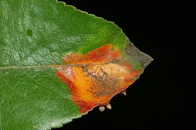
The subphylum Pucciniomycotina consists of a variety of fungi including yeasts, jelly fungi, rusts, and smut. However, rusts are some of the most common members of the group and will be discussed in detail.
Some examples of Pucciniomycotina species in phylum Basidiomycota include:
- Mixia osmundae
- Microbotryum violaceum
- Microbotryum dianthorum
- Cystobasidium psychroaquaticum
- Cystobasidium fimetarium
- Occultifur externus
- Occultifur tropicalis
Life Cycle and Morphology of Rust
The majority of Pucciniomycotina (over 7,000 species) are in the rust lineage. They have a complex life cycle that involves several spore stages depending on the species. To understand the life cycle and morphology of rust fungi, this section will focus on the fungus Puccinia graminis.
Compared to some of the other fungi (e.g. most mushrooms), the fungus Puccinia graminis is an obligate parasite and therefore requires a host (two hosts/heteroecious) for survival.
The following are some of the main steps in the lifecycle of this fungus:
Stage 1: Uredinial stage (Uredospores)
In this stage, the aeciospore lands on the plant (wheat) leaf or stem. Upon landing, it produces a germ tube that grows along the epidermis to reach the stomata. Upon reaching the stomata, the tip of the germ tube swells to form the appressorium. Contents of the germ tube (cytoplasm and nuclei) migrate to the vesicle (appressorium) and a septum forms separating it from the germ tube.
Over time, the appressorium produces a hypha (penetration hypha) that allows the organism to enter the plant through the stomata. Within the sub-stromal chamber, the tip swells to form a substromal vesicle in which contents of the appressorium are transported.
The vesicle gives rise to dikaryon hypha (with two nuclei in each cell) also known as infection hypha. In turn, the hypha branches and gives rise to hyphae threads that grow and spread between the cells within the plant cell/stem.
As the mycelium obtain nutrients between the cells, they cluster and give rise to short, upright hyphae known as uredinia/uredia. Urediniospores are released from the tips of these bodies in large numbers causing the epidermis to rupture.
* Also known as uredospores, the dikaryotic erediniospores are generally oval in shape with a stalk. They also have a thick wall and are reddish in color. If they are dispersed and transported by the wind, they can fall onto new hosts and germinate.
Stage 2: Telial stage
This part of the life cycle occurs at the end of the host plant (wheat) growing season when it becomes hotter and drier. During this period, mycelia give rise to telia that produce spores known as teliospores/teleutospores. These spores contain two cells and have a thick, black smooth wall.
These spores can survive for a long period of time and eventually germinate during low temperatures.
Stage 3: Basidial stage (basidiospores)
In the third stage, teleutospores produce thin hyphae known as promycelium. Along with the teleutospore cell, the promycelium is known as a basidium. From the teleutospore, the diploid nucleus moves to the promycelium where they undergo meiotic division to produce four haploid nuclei and eventually four haploid cells.
Each of the cells then produces thin, tube-like structures known as sterigma. At the tip, the sterigma form spore-like cells into which contents of the promycelium migrate (including the haploid nucleus). This results in the formation of spores known as basidiospores. Generally, these spores are colorless and have a thin wall. Unlike previous spores, these spores cannot infect cereal hosts.
Stage 4: Pycnidial stage
Basidiospores formed in the previous stage are transported to the alternative host (Barberry plant) by the wind. Here, they produce haploid mycelia that penetrate the epidermis of the host leaf and produce spermogonia (pycnia).
Characterized by a flask-like morphology, these structures (pycnia) produce haploid gametes (spermogonia and receptive hyphae).
Spermogonia (spermatia) gametes are produced at the tips of short, hyphae located at the base of spermogonium. Here, they are released in large numbers from the ostiole in a sticky substance that attracts insects and allows for transportation.
Receptive hyphae, on the other hand, are produced in the neck of pycnium and grow out through the ostiole. Unlike spermatia, these hyphae (they might be branched) are female gametes.
* Fertilization of the receptive hyphae by spermatia produces dikaryotic mycelium. This is a form of sexual reproduction.
Stage 5: Aecidial stage
In the last phase of Puccinia life cycle, the dikaryotic mycelium grows into the tissue of the leaf where they form aecidio mother cells. These structures in turn produce dikaryotic spores known as aecidiospores characterized by a hexagonal morphology.
The chain of aecidiospores is surrounded by the aecidial cup which emerges at the surface of the leaf. These spores are dispersed by the wind and only germinate when they land on cereal hosts (wheat). Here, they produce germ tubes that allow them to penetrate the plant and the cycle repeats itself.
* The rusts vary in color depending on the species.
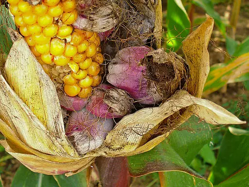
* Smuts are multicellular fungi that produce thick-walled spores. They are parasites of various plants including flowers, and are characterized by dark, soot-like masses known as sori.
* While some of the species are parasites of plants, some, (yeasts) can be found in association with other organisms like mosses and some vascular plants.
Yeast species can also be found in marine and, terrestrial freshwater habitats.
Ustilaginomycotina
The subphylum Ustilaginomycotina consists of over 1,700 species most of which are parasites of plants. Some of the species in the group have been shown to parasitize mammals.
Unlike the two other sub-divisions, Ustilaginomycotina does not contain mushroom fungi.
Here, the majority of species are smut fungi and cause diseases to a variety of plants including maize, wheat, and sugarcane. In their life cycle as dimorphic parasites, they go through the haploid stage and a dikaryotic hyphal stage.
The haploid stage involves the fusion of sexually-compatible yeast-like cells, the hyphal stage is initiated.
Generally, the life cycle may be represented as follows:
Haploid stage - Haploid saprobic yeast forms mate (plasmogamy) to form hyphal forms. Here, cells of the hyphae contain a pair of nuclei.
Infection - Hyphae infect the plant and produce spores (teliospores). Teliospores germinate and give rise to the basidium.
Zygote - Basidium gives rise to a zygote through a process known as karyogamy.
Haploid yeast forms - The zygote undergoes meiosis and give rise to the saprobic yeast forms and the cycle repeats itself.
Some species of the Subphylum Ustilaginomycotina in phylum Basidiomycota include:
- Ustilago tritici - parasite of rye, barley, and wheat
- Ustilago maydis - Parasite of corn/maize
- Ustilago spegazzinii - Parasitize twitch grass
- Exobasidium myrtilli - Parasite of some flowers
Phylum Basidiomycota Sexual Reproduction/Development
The majority of fungi are capable of sexual and asexual reproduction. Among members of the phylum Basidiomycota, sexual reproduction largely involves the fusion of haploid yeast cells (in yeast forms) or homokaryotic hyphae. This process gives rise to dikaryon forms with two nuclei (one from the haploid forms). The nuclei are replicated resulting in the elongation of the hyphae.
Asexual reproduction, on the other hand, occurs through the formation of asexual spores or budding. Here, some of the hyphae develop to produce asexual spores that germinate and give rise to a mature fungus.
Budding is different in that it involves the outgrowth of the parent cell to produce a daughter cell that grows and matures as the cycle continues.
What does Phylum mean in Biology?
Return from phylum Basidiomycota to MicroscopeMaster home
References
David S. Hibbet. (2006). A phylogenetic overview of the Agaricomycotina.
Jason E. Stajich. (2009). The Fungi.
JohnWebster and Roland Weber. (2007). Introduction to Fungi.
Marco A Coelho, Guus Bakkeren, Sheng Sun, and Michael E Hood. (2017). Fungal Sex: The Basidiomycota.
Links
https://steemit.com/puccinia/@mameen745/life-cycle-of-puccinia-graminis-best-article
https://www.sciencedirect.com/topics/agricultural-and-biological-sciences/basidiospore
https://www.researchgate.net/publication/267810920_Pucciniomycotina
Find out how to advertise on MicroscopeMaster!


