Phylum Apicomplexa
Definition, Classification and Characteristics
Definition: What is the Apicomplexa?
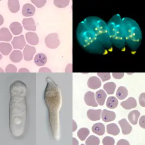
Previously called Protozoa, along with several other groups, the Phylum Apicomplexa is large and is further divided into 300 genera and over 60 families that consist of over 5000 species. The majority of species already identified are obligate intracellular parasites that infect a variety of animals (including human beings) causing a variety of diseases.
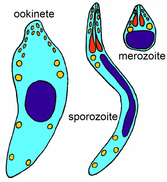
Because of the high diversity in this group, members have also been shown to exhibit a wide range of morphological shapes and characteristics depending on the genus and stage in their life cycle. This diversity also extends to the environment/habitat in which they are found in nature.
* Despite the high diversity in shape, characteristics, and where they are found in nature, members of this group are characterized by the apical complex and apicoplast.
Common members of this group include:
- Babesia
- Plasmodium
- Eimeria
- Isospora
- Haemoproteus
- Haemogregarina
Classification of Phylum Apicomplexa
Superkingdom: Eukaryota - Includes members with a membrane-bound nucleus
Kingdom: Chromalveolata (2005 classification) - A large group consisting of morphologically variable protists. The group is divided into four main groups that include Heterokontae, Alveolatae, Hacrobiae, and Rhizariae.
Infrakingdom/superphylum: Alveolata - Members of this group are single-celled eukaryotes that are widely distributed in different environments/habitats. They are characterized by a diverse means of locomotion as well as flattened vesicles known as alveoli. Apart from apicomplexans, other organisms classified in this group include ciliates and dinoflagellates.
Phylum: Apicomplexa
The phylum is further divided into two classes that include:
- Aconoidasida
- Conoidasida
Aconoidasida
Created in 1980, Aconoidasida is one of the two main classes of apicomplexan parasites.
It consists of members of the family Babesiidae and Theileriidae and has the following characteristics:
- Do not possess conoid in their apical complex
- Reproduce asexually through multiple fission
- Do not have specialized structures for locomotion
- Their oocysts do not have a cyst wall
Conoidasida
The class Conoidasida was introduced by Levine in 1988. It's further divided into subclasses coccidia (mostly infect vertebrates) and gregarines (parasites of invertebrates) which are characterized by a hollow conoid. They reproduce through sexual and asexual means and produce flagellated microgametes. Although some of the species have pseudopodia, they are mostly used for phagocytosis and not for movement.
Distribution
As mentioned, the majority of already identified phylum Apicomplexa species are parasites of animals and human beings. For this reason, their distribution across the world is largely dependent on the species, environmental conditions (climate), and the host they infect.
Malaria parasites (plasmodium species) are largely restricted to tropical and subtropical areas as well as in altitudes below 1,500m where they are responsible for high cases of malaria. On the other hand, B. divergens, a species of the Genus Babesia, is largely distributed in Europe with many cases (infections) being in human beings.
Depending on the type of host infected, Apicomplexa species are also found in different environments across the world. Moreover, the concentration of given species will vary from one environment to another.
In a study to investigate the distribution of coccidian parasites in aquatic and terrestrial environments in infected snakes, studies found a variation in infections between strictly terrestrial (52 percent) and semi-aquatic snakes (48 percent). This variation extends to different regions across the world.
* Apicomplexa and related parasites can be found in marine and freshwater environments where they infect such hosts as Solea senegalensis.
Characteristics
Cell Structure and Morphology
Apicomplexans are eukaryotic organisms and are therefore characterized by a complex structure as compared to the structure of prokaryotes. As eukaryotic organisms, they have membranous organelles (e.g. nucleus, endoplasmic reticulum, and mitochondria, etc).
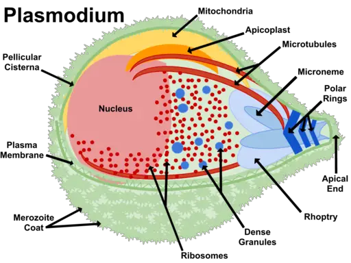
They are also highly polarized cells that contain a given set of organelles that are only found in the phylum. They contain such secretory organelles as rhoptries and micronemes with given products that contribute to motility and cell invasion.
Flagella, used for motion, are commonly found in the gametes (male gametes). However, the number of flagella varies from one species to another. While male gametes in Plasmodium contain a single flagellum, Toxoplasma microgametes are bi-flagellated and thus contain two flagella. In both cases, the structure consists of 9 doublet microtubules and a central pair.
* There are two types/categories of microtubules in apicomplexans. These include subpellicular microtubules (responsible for the shape and polarity of the apical complex) and spindle microtubules (involved in nuclear division during mitosis).
Apical Complex
The apical complex is the defining structure of apicomplexans. Located at the anterior end of adult obligate parasites, the apical complex consists of cytoskeleton structures and membrane-bound organelles.
The primary components of this structure include:
The conoid - The conoid is a cytoskeletal structure found in some of the apicomplexan parasites. It can be found in Toxoplasma and Eimeria species among others but is absent in such apicomplexans as Theileria and Plasmodium.
Essentially, the conoid is composed of counterclockwise spiraling filaments that form the cone-shaped structure at the apex of these apicomplexans. Based on molecular studies, filamentous subunits of the conoid have been shown to resemble microtubules. However, they are curled into a coil. They are about 250nm in diameter with two 400nm long microtubules located in the middle.
Rhoptry - Rhoptries are membrane-bound organelles (twin) with a pear-shaped appearance. They are part of the apical complex that secretes proteins at the apical tip.
Also described as secretory lysosomal granules, rhoptries (between 8 to12 per cell) also receive material from biosynthetic and endocytic cell pathways - Rhoptries are also associated with micronemes and dense granules in the parasite.
Polar rings - Found in all apicomplexans, polar rings serve as microtubule-organizing centers for the subpellicular microtubules.
Apicoplast
Discovered in the 1970s, the apicoplast is an organelle of apicomplexans that contains its own DNA (as is the case with other plastids). Because they are non-photosynthetic plastids, apicoplasts are mostly regarded as vestigial plastids of apicomplexan parasites. They are suggested to have originated from endosymbiosis of cyanobacteria about 1 billion years ago.
As plastids, the apicoplast contains 35kb circular genome. Today, many of the studies focusing on the organelles are aimed at targeting it for treatment purposes.
Life Cycle and Reproduction
Members of the phylum Apicomplexa have a complex life cycle that is characterized by three major processes that include merogony, sporogony, and gametogony. For most species, this cycle alternates between sexual and asexual stages in one or different hosts depending on the organism. For different species, the cycle may vary slightly with different terminologies being used to describe the different stages.
In this section, reproduction and life cycle of Toxoplasma gondii will be used to represent the group:
Like many other members of the phylum Apicomplexa, Toxoplasma gondii is a very successful parasite. It's widely distributed in many parts of the world (from the very cold arctic to deserts) where it infects a variety of hosts (intermediate and definitive hosts).
* Whereas domestic cats and other felids act as the natural hosts of Toxoplasma gondii, non-feline species serve as the intermediate hosts.
The sexual cycle of reproduction occurs in the natural (definitive) host. This starts when a cat (or any other felid) ingests infected tissues (consisting of bradyzoite cysts) or oocysts. For most cats, this happens when they feed on infected intermediate hosts (e.g. birds). Once the infected prey is ingested, along with tissue cysts, bradyzoites are released in the stomach and intestines from the cyst.
Here, the digestive enzymes in the intestine/stomach act on the cyst causing it to dissolve and release the bradyzoites. Once they are released, the bradyzoites then enter the epithelial cells of the small intestine where they give rise to schizonts.
Here, the schizonts undergo several stages of asexual division to release merozoites that ultimately produce the male and female gamonts. Following the union of these cells, fertilization takes place forming the macrogamont that is then covered by a wall to form the oocyst.
This phase in the life cycle of the parasite is known as enteroepithelial life cycle and is limited to the feline hosts. The different parasitic forms formed during this phase have different morphological characteristics that allow them to thrive.
Bradyzoites - Bradyzoites measure between 3 and 4 by 1 to 2um in size. Before they are released, they are covered by a thin cyst wall that protects them from the external environment. They are resistant to the digestive enzymes which allow them to survive in the intestine of the host.
Schizonts - Schizonts are small intracellular bodies that divide rapidly. They measure 4 to 5 by 1 to 2um in size. Unlike bradyzoites, schizonts normally develop in the vacuoles of the epithelial cells.
Merozoites - Merozoites represent the motile stage in various organisms. As such, they are capable of moving from one cell to another thus causing significant cell destruction. Because of their ability to divide rapidly, they contribute to the large population of the parasite.
Oocysts - May appear round or oval and measure about 10 by 12 um in size.
The oocysts formed are then released into the environment along with fecal matter. At this stage, they are unsporulated given that they are contained in a thin wall.
Following a period of between 1 to 5 days, the start to sporulate. According to studies, this is influenced by environmental conditions (air and moisture etc). The sporulation process results in the production of sporocysts (two) each of which contains four sporozoites.
Once the oocysts are ingested by the intermediate host, the sporozoites hatch in the lumen in the small intestine allowing the sporozoites to be released and enter the intestinal cells. Here, the cells undergo endodyogeny (asexual process) and give rise to tachyzoites.
Tachyzoites are rapidly dividing cells capable of dividing within any cells in the body. Because of the rate at which they divide, they cause cell destruction and infect new cells causing even more damage.
At some point, they encyst to form tissue cysts that vary in size from about 15um to 60um. They are commonly formed in the central nervous system, muscles, and visceral organs. In these sites, they can persist for a long time (until the host dies). In the event that this host is consumed by a cat, then the cycle continues.
Host Cell Invasion
Apicomplexans are obligate intracellular parasites that invade and divided within the cells of the host.
This is a stepwise process that can be divided into four main phases that include:
Host cell approach - At this stage, most parasites are capable of motility. This is mostly achieved by gliding which is made possible by a number of components including actin, myosin A motor complex, as well as micronemal transmembrane proteins. This type of motility allows the cell to move without any deformation and is at the rate of a few microns per second.
Host cell recognition - In order to successfully invade the cell, apicomplexans have to identify the cell to be invaded. It allows the parasite to invade the appropriate cell with favorable conditions for multiplication. Plasmodium parasites invade red blood cells that provide conducive conditions for division. Here, micronemal proteins have been shown to play an important role in the recognition of the host cell as well as attachment to the cell.
Formation of a tight junction and entry - The formation of a junction between the parasite and the cell of the host is an important step that allows the parasite to penetrate the cell. Following contact (between the cell of the host and the parasite), rhoptries, which are apical secretory organelles, secrete and inject their contents (proteins capable of altering the functions of the host cells) into a vacuole and consequently into the cytosol of the host cell.
This results in the formation of evacuoles (small vesicles). It creates a junction between the cell and the parasite allowing the parasite to penetrate the cell. Here, the gliding action of the parasite has been shown to promote entry.
* Once the parasite penetrates the cell, it can enter the vacuole where it can develop and divide for multiplication purposes or in order to give rise to a new form of the parasite.
Some of the diseases caused by those in the phylum Apicomplexa include:
- Malaria - Caused by Plasmodium species
- Toxoplasmosis - Caused by Toxoplasma gondii
- Coccidiosis - Caused by Eimeria tenella
- Babesiosis - Caused by Babesia
- Cyclosporiasis - Caused by Cyclospora cayetanensis
What does Phylum mean in Biology?
Return from phylum Apicomplexa to MicroscopeMaster home
References
Fernando Martínez-Ocampo. (2017). Genomics of Apicomplexa.
L. David Sibley. (2010). How Apicomplexan Parasites Move In and Out of Cells.
Maria E. Francia, Jean-Francois Dubremetz & Naomi S. Morrissette. (2015). Basal body structure and composition in the apicomplexans Toxoplasma and Plasmodium.
Naomi Morrissette and L. David Sibley. (2002). Cytoskeleton of Apicomplexan Parasites.
Links
http://parasite.org.au/para-site/text/toxoplasma-text.html
Find out how to advertise on MicroscopeMaster!


