Phase Contrast Microscope
Buyer's Guide; Application; Advantages and Disadvantages
The Phase Contrast Microscope opened up an entire new world in the field of microscopy.
Up until this discovery, scientists were limited to bright field illumination and did not have the ability to view live microorganisms.

Now, phase contrast observation is a standard feature on almost all modern microscopes.
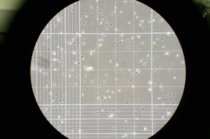
To the right is a view through the eyepiece of a standard phase contrast microscope showing a hemocytometer with fibroblasts.
History and Background Information
Frits Zernike, a Dutch physicist and mathematician, built the first phase contrast microscope in 1938.
It took some time before the scientific community recognized the potential of Zernike’s discovery; he won the Nobel Prize in 1953 and the German-based company Zeiss began manufacturing his phase contrast microscope during World War II.
Zernike experimented with the speed of the light path directed onto a specimen; he discovered interference patterns resulting in the image appearing darker or lighter.
He developed a system using annuli or rings, placed in the lens and beneath the lower condenser of a compound microscope, to cause interference in light patterns. Zernike manipulated the rings and light source, ultimately reducing the light wavelength by a ½ phase. Each magnification setting, whether 10x or 100x, needs an analogous annulus ring in the light condenser.
The quality of observation, specifically the contrast and resolution of a specimen or particle (P), is contingent on the relationship between the surround (S) waves (also known as un-diffracted or zeroth-order) and diffracted (D) spherical wavefronts. The primary equation behind phase contrast is P=S+D.
This process enabled him to separate zeroth-order (S) from diffracted light (D), increasing the definition and visibility of images/particles (P).
In essence, Zernike’s approach and adjustments to illumination changed the way we use microscopes and view specimens.
Phase Objects
Phase contrast is most useful in observing transparent, colorless and/or unstained specimens referred to as “phase objects.”
Since these objects do not absorb light, they could not be seen with any detail before Zernike’s discovery.
The new system took advantage of both direct and diffracted light to increase the quality and definition of transparent samples.
Most importantly, many phase objects are living biological samples; the potential implications and uses of phase contrast within the world of microscopy should not be undervalued.
Today, two main types of phase contrast are positive and negative. Since the observed particles are usually thin and transparent, these polar contrasts provide strikingly different images.
Positive phase contrast reveals medium to dark gray images on a lighter grey background; these images often have a bright halo along the edge of the sample.
Negative phase contrast is the opposite. The specimen appears lighter with a dark background; they also have a dark halo outlining the image.
Applications in Microscopy
The possible applications of Zernike’s phase contrast microscope in microscopy are evident in the fields of molecular and cellular biology, microbiology and medical research.
Specimens that can be observed and studied include live microorganisms such as protozoa, erythrocytes, bacteria, molds and sperm, thin tissue slices, lithographic patterns, fibers, glass fragments and sub-cellular particles such as nuclei and organelles.
Advantages
The advantages of the phase contrast microscope include:
- The capacity to observe living cells and, as such, the ability to examine cells in a natural state
- Observing a living organism in its natural state and/or environment can provide far more information than specimens that need to be killed, fixed or stain to view under a microscope
- High-contrast, high-resolution images
- Ideal for studying and interpreting thin specimens
- Ability to combine with other means of observation, such as fluorescence
- Modern phase contrast microscopes, with CCD or CMOS computer devices, can capture photo and/or video images
In addition, advances to the phase contrast microscope, especially those that incorporate technology, enable a scientist to hone in on minute internal structures of a particle and can even detect a mere small number of protein molecules.
Disadvantages
Disadvantages and limitations of phase contrast:
- Annuli or rings limit the aperture to some extent, which decreases resolution
- This method of observation is not ideal for thick organisms or particles
- Thick specimens can appear distorted
- Images may appear grey or green, if white or green lights are used, respectively, resulting in poor photomicrography
- Shade-off and halo effect, referred to a phase artifacts
- Shade-off occurs with larger particles, results in a steady reduction of contrast moving from the center of the object toward its edges
- Halo effect, where images are often surrounded by bright areas, which obscure details along the perimeter of the specimen
Modern advances and techniques provide solutions to some of these confines, such as the halo effect.
Apodized phase contrast utilizes amplitude filters that contain neutral density films to minimize the halo effect. Essentially, this is attempting to reverse the definition achieved through phase contrast annuli, but the halo effect can never be eliminated completely.
The pros that phase contrast has brought to the field of microscopy far exceed its limitations. This is easily seen with the myriad of advances in the fields of cellular and microbiology as well as in medical and veterinary sciences.
MicroscopeMaster's Phase Contrast Microscope Reviews/Guides
Check out:
Omax 40X-2500X Phase Contrast Trinocular LED Compound Microscope + 9MP USB Camera
Omax 40X-2500X PLAN Infinity Phase Contrast Trinocular Siedentopf Microscope
AmScope T490A-PCT Compound Trinocular Microscope with Phase Contrast turret

Conclusion
The phase contrast microscope opened up an entire world of microscopy, providing incredible definition and clarity of particles never seen before.
These transparent specimens could not be explored because they do not have the capacity to absorb light.
Zernike found a way to manipulate light paths through the use of strategically placed rings and his system is a staple of most modern microscopes.
Although there are a few disadvantages, such as shade-off and halo distortions, phase contrast provides highly detailed, well-contrasted images.
The most important breakthrough is the ability to observe living particles in a natural state.
For further information about techniques available, go ahead and read about other microscopy imaging techniques.
Of interest: Phase Contrast Microscopy Images
Return to Compound Light Microscope
Return from Phase Contrast Microscope to Best Microscope Home
Find out how to advertise on MicroscopeMaster!


