Peroxisomes
An Overview - Definition, Function and Structure
Introduction/Overview
|
First detected in the 1950s, peroxisomes are small ubiquitous organelles found in virtually all eukaryotic cells. Unlike many of the other organelles that serve one or a few functions, peroxisomes have been associated with diverse functions in different organisms ranging from biosynthesis of penicillin in fungi to various metabolic reactions in mammals. They have also been associated with a variety of other functions including signaling, aging, as well as a role in immunity thus making them essential cell organelles. |
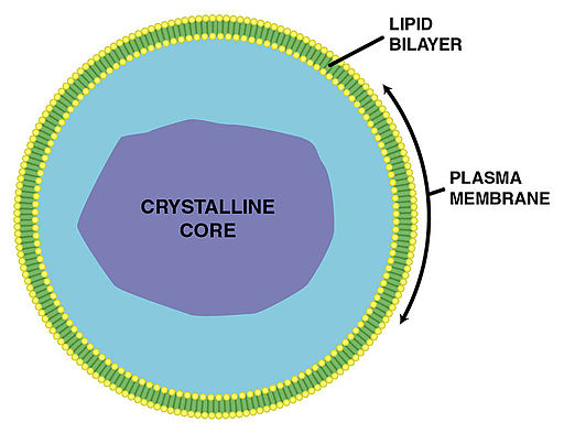
In mammalian cells, they are estimated to occupy about 2 percent of the total cell volume and are characterized by a granular matrix and a single membrane. In different organisms, peroxisomes have diverse names depending on the substance they produce and their functions.
These include:
· Glycosomes - involved in glycolytic reactions in trypanosomatids
· Glyoxysomes - contain enzymes involved in the glyoxylate cycle in plants
· Woronin body - found in filamentous fungi where they are involved in sealing the septal pore and thus contribute to cellular integrity
* Peroxisomes were first identified by Johannes Rhodin in 1954 in mice (he called them microbodies). It was not until 1965 that Christian de Duve proposed the name Peroxisomes.
* Peroxisomes are important for cellular homeostasis, vitality, and proper development of an organism. Disorders of peroximoses have been associated with such conditions as the Zellweger syndrome and neonatal adrenoleukodystrophy among other disorders collectively known as peroxisome biogenesis disorders.
Origin of Peroxisomes
Based on various characteristics of the organelle, several hypotheses have been presented to explain the origin. One of these hypotheses suggests that peroxisome is the result of an endosymbiotic relationship involving bacteria. However, because of the apparent similarity between some of the proteins of the organelle and those found in the endoplasmic reticulum, some researchers are of the idea that they developed from the endoplasmic reticulum.
Regardless, evolution of these organelles from a common ancestor has become widely accepted for a number of reasons.
Despite having different functions and even variation in size, etc, the core mechanism by which division, biogenesis, and maintenance occur in peroxisomes is the same. Most of the new evidence, however, supports the hypothesis that their origin is related to the endoplasmic reticulum.
In their development, studies have shown that some of the peroxisomes membrane proteins first target the endoplasmic reticulum before they reach the peroxisomes. In addition, new peroxisomes have been shown to form from the endoplasmic reticulum following the introduction of the wild-type gene in yeast.
Morphology and Structural Characteristics
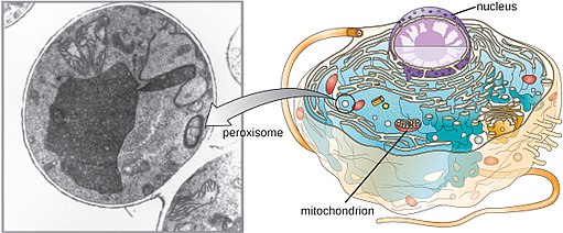
Also referred to as microbodies in some books, peroxisomes are very small in size, ranging from 0.2 to 1.5 um in diameter. While the size varies between different organisms (mammals, plants, fungi, etc), studies have shown them to also vary in size within the same organism.
A single cell may also contain numerous peroxisomes depending on the organism. In mammals, for instance, a single hepatocyte (liver cell) may consist of between 400 and 600 peroxisomes that occupy about 2 percent of the cell volume.
Generally, peroxisomes are spherical in shape and contain a single membrane of between 4.5 and 8 nm in diameter. This membrane is made up of a number of components including phospholipids—phosphatidylcholine, phosphatidylethanolamine, and phosphatidylinositol. It's thinner when compared to the membrane of other organelles (e.g. lysosomes etc). However, the thickness is similar to that of the endoplasmic reticulum.
Like the plasma membrane, the peroxisome membrane is also permeable and can allow such molecules as sucrose and other smaller substrates to pass through. This permeability, however, may vary depending on the cell location.
A marginal plate has also been described in the Peroxisomes of some species. Here, the marginal plate tends to be flat and is typically located at the periphery of the organelle where it lies within a narrow space and surrounded by the matrix. As such, it's separated from the inner surface of the surrounding membrane.
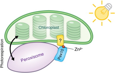
* In the cell, peroxisomes can be found floating in the cytoplasm. However, they often include association with a number of other organelles including the mitochondria, chloroplast (in plants) as well as the endoplasmic reticulum.
* They may be interconnected to form the peroxisome reticulum of simply exist as individuals and contain a matrix (made up of fibrils/crystalloid material).
Biogenesis and Division of Peroxisomes
The biogenesis of peroxisomes is a relatively complex process that involves several phases that include the formation of the peroxisome membrane, import of matrix proteins as proliferation. While the process is not fully understood, studies have shown that the biogenesis process starts with the development of the membrane.
Here, proteins known as peroxins (PEX - including PEX3, PEX16, and PEX19) serve to insert membrane proteins (PMPS - peroxisomal membrane proteins) into the membrane of the organelle. In mammals, the endoplasmic reticulum is also suggested to play an important role in supplying the necessary lipids required for the development of the membrane.
* While lipids are recruited from the endoplasmic reticulum, proteins used during the biogenesis of peroxisomes are produced in the polyribosomes that reside in the cytoplasm.
Following the processes involved in biogenesis, peroxisomes continue to multiply through division (fission). For different organisms, a number of factors influence division of peroxisomes in cells. In yeast, for instance. Studies have shown this to be influenced by the level of protein matrix.
Following the sequestration of the peroxisomal membrane protein by fatty acyl-CoA oxidase (an enzyme located in the matrix), a series of events are activated resulting in the production of diacylglycerol. In turn, the diacylglycerol causes membrane curvature and consequently the recruitment of division factors.
During peroxisome division, the organelle elongates and constricts at several parts in a manner that produces divisible units. The last phase of this process involves the division of the units (fission) to produce several peroxisomes.
* The division process may be symmetrical or asymmetrical. In the case of symmetrical division, the process produces several peroxisomes of the same size. On the other hand, asymmetrical division results in the production of peroxisomes of varying sizes.
* Following division and multiplication of the peroxisomes, they move to different locations of the cell before the cell divides to produce two similar daughter cells. This movement is made possible by microtubules. Here, the peroxisomes travel along these tubules allowing them to be partitioned into the daughter cells in roughly equal numbers.
Characteristics (common traits)
Despite the differences in size and functions, all peroxisomes share a number of characteristics. One of these characteristics is the presence of a single membrane that surrounds the entire structure. In all peroxisomes, the lumen contains a high amount of enzymes that are involved in various functions.
These functions are largely dependent on the type of organism as well as the type of tissue. Unlike some of the other organelles found in a cell (mitochondria and chloroplasts etc), peroxisomes lack their own genome. For this reason, the processes involved in peroxisomes division are regulated by the genetic material of the cell (peroxisomal proteins are encoded by the nuclear genome of the cell).
One of the other similarities shared between all peroxisomes is the mechanism through which the membrane is made as well as the process involved in division/fission.
According to research studies, proteins and lipids involved in the creation of the peroxisomal membrane are imported. Whereas the peroxisomal proteins are manufactured by free ribosomes in the cytoplasm and transported to the structure, the lipids are imported from the endoplasmic reticulum.
As well, the division process in all peroxisomes involves the activities of the dynamin-like protein and a protein that contains the Tetratrico Peptide Repeat.
Main Functions
As mentioned, peroxisomes have diverse function in plants and animals.
Some of the main functions of these organelles include:
Photorespiration
Photorespiration is one of the main function of peroxisomes in plants. This is an important process that is associated with photosynthesis and involves the activities of RubisCo. Here, the process starts with the molecule (RubisCO) taking oxygen as a substrate. This results in the production of phosphoglycolate which then undergoes dephosphorylation to produce glycolate.
In the matrix of the peroxisome, the glycolate is oxydated by glycolate oxydase producing glycoxylate as well as hydrogen peroxide (H2O2). Two enzymes (SGT and GGT) then act on the glyoxylate in a process known as transmination to produce glycine.
In the mitochondria, glycine is converted to serine which is then transported back to the peroxisomes where it is converted to glycerate and hydroxypyruvate. The glycerate is then transported to the chloroplast where it is involved in the formation of glucome.
* Photorespiration is an important process involved in the recycle of carbon.
Oxidation of Fatty Acids
One of the other functions of peroxisomes in plants involves the degradation of fatty acids. For this to take place, fatty acids are first transported to the peroxisomes and transformed into the CoA-esters, a form that enters the β-oxidation cycle.
This process is particularly important in that it converts the long fatty acid chains (with 20 or more carbon atoms) into acetyl CoA which is a source of metabolic energy. This process also produces hydrogen peroxide which is broken down by catalase to produce oxygen and water molecules.
Some of the other important functions of peroxisomes in plants include:
· The biosynthesis of jasmonate - Jasmonate is a group of hormones (e.g. methyl jasmonate) involved in plant growth and development as well as defense mechanisms
· Metabolism of Indole-3-butyric Acid
· Metabolism of polyamine
· Metabolism of branched-amino acid chain
· Seed germination
Detoxification
In both plants and animals, peroxisomes are heavily involved in detoxification. Through the various metabolic functions of this organelle, hydrogen peroxide is one of the by-products produced. In the body, this product is harmful and can interfere with other cellular functions.
Also, peroxisomes produce high amounts of catalase, an enzyme that breaks down this chemical into water and oxygen molecules. The peroxide can be used for the oxidation of organic compounds (those that contain carbon).
* Signaling - Peroxisomes have also been shown to play an important in immunity. Here, the organelles produce bioactive metabolites involved in immune signaling. In various studies, these processes have been associated with antiviral responses in animals.
Peroxisomes and Cell Aging
Generally, it's well understood that various activities within the mitochondria result in the production of reactive oxygen species that exert significant stress on living cells. In the process, important cellular components (lipids, proteins etc) are somewhat damaged which causes cell deterioration over time as well as a loss of cell viability over time.
Through research studies to understand biological functions of peroxisomes, it has become evident that like mitochondria, peroxisomes also produce a significant level of reactive oxygen species that contribute to cell aging.
According to a study that was aimed at investigating the relationship between peroxisomes and cell aging (in yeast cells), researchers noticed that in cells with a relatively low level of hydrogen peroxide (a reactive oxygen species) cells that lacked catalase (an enzyme responsible for breaking down hydrogen peroxide), the cells had a longer life span compared to the wild-type cells (natural cells) used as the control.
In cells where high amounts of the reactive oxygen species was produced, cells without catalase had a significantly shorter life span compared to control cells. It became evident that the reactive oxygen species produced through metabolic activities in the peroxisomes not only cause damage to various macromolecules of the cell, but also contribute to cell damage and loss of viability.
This study showed the significant role of catalase which breaks down the peroxide thus acting as an anti-oxidant that would otherwise accelerate cell aging and cell death.
Diseases/Disorders associated with Peroxisomes
Disorders of peroxisomes, as well as defects in the production of peroxisome enzymes, have been associated with a number of peroxisomal diseases that include:
· Zellweger syndrome - A disorder resulting from reduced/absence of properly functioning peroxisomes. It's characterized by hypotonia, hearing loss, a loss of vision, as well as skeletal abnormalities and distinct facial features
· Neonatal adrenoleukodystrophy - Results from defects in the biogenesis of peroxisomes or poor functioning peroxisomes. It may be characterized by hearing loss, seizures, hypotonia, and diffuse encephalopathy, etc
· Rhizomelic chondrodysplasia punctata - A disorder characterized by seizures, infections of the respiratory tract, and rhizomelia
· Infantile Refsum's disease - An inherited disorder characterized by damage to the white matter of the brain - This also affects motor movements
Return from learning about Peroxisomes to MicroscopeMaster home
References
Francesca Di Cara et al. (2019). Peroxisomes in Immune Response and Inflammation.
Marten Veenhuis and Ida J. van der Klei. (2002). Peroxisomes: surprisingly versatile organelles.
Stanley R. Terleckya, Jay I. Koepkea, and Paul A. Walton. (2006). Peroxisomes and aging.
Selvambigai Manivannan, Christian Quintus Scheckhuber, Marten Veenhuis and Ida Johanna van der Klei. (2012). The impact of peroxisomes on cellular aging and death.
Toni Gabaldón. (2010). Peroxisome diversity and evolution.
Links
https://bscb.org/learning-resources/softcell-e-learning/peroxisome/
https://mmegias.webs.uvigo.es/02-english/5-celulas/6-peroxisomas.php
https://www.researchgate.net/publication/221803946_Peroxisome_Biogenesis_and_Function
Find out how to advertise on MicroscopeMaster!


