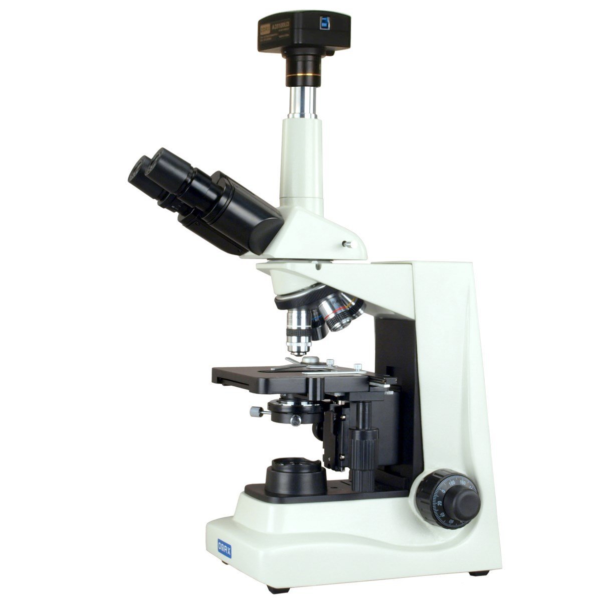
OMAX 40X-2000X USB3 14MP PLAN Trinocular
Darkfield Lab Microscope
Super Bright LED for Live Blood
Overview
|
The OMAX 40X-2000X USB3 14MP PLAN Trinocular Darkfield Super Bright LED Lab Microscope is one of the best microscopes for live blood analysis. It is an excellent device that comes fully equipped with a number of great features that make it the perfect microscope for research laboratory applications and advanced applications in clinical offices. |
|
Main features
- A Trinocular Siedentopf head
- A diopter
- A double layer mechanical stage
- A digital camera
- Darkfield condenser
Product Description
Microscope Head
The USB3 14MP PLAN Trinocular Darkfield Super Bright LED Lab Microscope comes with a Trinocular Siedentopf, which is inclined at a 30 degrees angle and can rotate 360 degree. This is an important feature of the Siedentopf head in that it enhances comfort during use, allowing the user to turn the head and observe the specimen at their own comfort.
With a Siedentopf head in place, the interpupillary distance can also be changed without affecting focus, which is one of the biggest advantages of this microscope compared to sliding binoculars. It also comes equipped with a diopter on the left ocular tube, which can be adjusted to meet individual user's sight. As such, it can be easily used by more than one user without any problems regardless of differences in their eye vision. To switch between the ocular and photo tube, users only have to pull-out the bar, which is a very simple task.
Condenser
The OMAX USB3 14MP PLAN Trinocular microscope has in place an Abbe condenser NA1.25, a rack and pinion adjustment, with iris diaphragm and a filter holder. These are standard features that regulate the amount of light, ensuring that the user adjusts to just the right amount of light for clarity.
Digital Camera
The microscope comes with USB3 Digital camera, which is one of the best, all round camera with capabilities of dealing with all aspects of microscopy through its excellent image resolution power. The true color camera with 0.5X lens allows for a larger field of view thereby allowing for the capture of a large field of the image. This is in addition to super speed connection and frame speeds of 6.2fps at 4096x3286, 54fps at 1024x822.
The software is compatible with Windows, Mac OS X and Linux operating systems, which basically means that it can be used with any other PC. With the user friendly software, users will find it easy to not only capture and record live videos, but also measure lengths, angles, areas and edit images.
Some of the other important aspects of the OMAX 40X-2000X USB3 14MP PLAN Trinocular Darkfield Super Bright LED Lab Microscope include:
Optics - The microscope allows for eight levels of magnification (40X, 80X, 100X, 200X, 400X, 800X, 1000X, 2000X). This is a large range with high total magnifications that allow the microscope to be used for observing a variety of specimen. The two sets of eyepieces include the Widefield WF10X/18 and Widefield WF20X, which can be interchanged depending on the needs of the user.
All the objectives are Plan Achromatic objectives, which means that the curvature of the lens has been corrected. As a result, these objectives allow for a crisp clear image. The advanced oil darkfield NA1.36-1.25 condenser and iris controlled 100X NA1.25-0.5 objective also provide superior light control in darkfield for the best observation of live blood.
Stage - For this microscope, the stage is a double layer mechanical stage, which can easily be moved back and forth and side to side using the knobs below the stage. The stage also has in place long clips for holding the slide in place during observation. The slide therefore does not have to be moved manually with the hand, which may be a tedious task for some users.
Illumination - For the OMAX 40X-2000X USB3 14MP PLAN Trinocular Darkfield Super Bright LED Lab Microscope, the super bright LED in place provides additional light intensity that contributes to the clarity of the darkfield images at higher magnifications. The 100X Plan iris controlled objective also offers additional light control that minimizes influence from external light. This allows for clearer images of the specimen.
The package also comes with:
- Software (CD)
- Power code
- A USB cable
- A solid sturdy metal frame
- A super bright 5W LED light source
Darkfield Microscopy and Live Blood
Darkfield microscopy refers to the type of microscopy where light from the source below the stage illuminates the specimen. Here, light from the source is gathered in the condenser, shaped in to a cone and the apex focused on to the plane with the specimen.
In darkfield microscopy, the condenser has been specifically designed in a manner that ensures that it forms a hollow cone of light which is different from bright field microscopy where the sample is illuminated with a full cone of light.
In addition, the lens in darkfield microscopy sits in a dark hollow of the cone while light travels around the objective lens, but does not enter the cone shaped areas. Once the sample has been placed on the stage it appears bright against a dark background, allowing it to stand out. This technique is particularly useful for viewing blood cells, bacteria, algae and hairline metal fractures among others.
Live blood can be viewed using darkfield microscopes. Here, a drop of blood is placed on a slide. A drop of distilled water may be added and covered using a cover slip in order to keep it from drying out. The slide is then viewed under high magnification. Here, the image can also be observed on a PC or monitor. The cells appear as dark bodies. Using this technique, technicians can be able to observe and even analyze the cells.
Here's more on Blood Smears.
Pros and Cons - OMAX 40X-2000X USB3 14MP PLAN Trinocular Darkfield Super Bright LED Lab Microscope
This microscope is a professional device that presents significant benefits in such professional fields as clinical applications. It has several advantages in that it comes with a powerful and fast camera, allows for high magnifications and provides high resolution images that are very clear. As such, it is increasingly being used for observing and analyzing of blood cells.
While it has been shown to be an excellent microscope for observing live cells, some users may find it difficult to effectively analyze blood cells using the darkfield technique, which is why there are still varying opinions about using darkfield technique for blood analysis in medicine.
MicroscopeMaster reviews blood microscopy here
Conclusion
 |
The OMAX 40X-2000X USB3 14MP PLAN Trinocular Darkfield Super Bright LED Lab Microscope comes with a number of features that make it one of the best microscopes for observing a wide range of specimen. It can be used for both bright field and darkfield techniques thereby allowing for many uses. With an excellent camera and features that allow for clear and highly resolved images, it is worth considering when making a purchase. |
Return to Omax Microscope Buyer's Guide or Trinocular Microscope Main Page.
Find out how to advertise on MicroscopeMaster!


