Nucleus
** Definition, Structure & Function, Cellular vs Atomic Nuclei
Definition: What is a nucleus?
The nucleus is a membrane-bound organelle that contains genetic material (DNA) of eukaryotic organisms. As such, it serves to maintain the integrity of the cell by facilitating transcription and replication processes.
It's the largest organelle inside the cell taking up about a tenth of the entire cell volume. This makes it one of the easiest organelles to identify under the microscope.
Some of the other main components of a nucleus include:
- Phospholipid bilayer membrane
- Nucleoplasm
- Nucleolus
- Chromatic
* Some eukaryotic cells lack a nucleus and are referred to as enucleate cells (e.g. erythrocytes) while others may have more than one nucleus (e.g. slime molds).
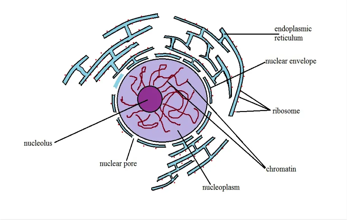
Diagrammatic Representation of a Nucleus
Structure and Organization of the Nucleus
As the organelle that contains the genetic material of a cell, the nucleus can be described as the command center. As such, the nucleus consists of a number of structured elements that allow it to perform its functions. This section gives focus to the structure of the cell.
In general, the nucleus has a spherical shape as shown in most books. However, it may appear flattened, ellipsoidal or irregular depending on the type of cell. For instance, the nucleus of columnar epithelium cells appears more elongated compared to those of other cells. The shape of a nucleus, however, may also change as the cell matures.
The Nuclear Membrane
The nuclear membrane is one of the aspects that distinguish eukaryotic cells from prokaryotic cells. Whereas eukaryotic cells have a nucleus bound membrane, this is not the case with prokaryotes (e.g. bacteria) that lack membrane-bound organelles.
As with the other cell organelles of eukaryotic organisms, the nucleus is a membrane-bound organelle. The nuclear membrane, like the cell membrane, is a double-layered structure that consists of phospholipids (forming the lipid bilayer nucleus envelope).
Present on the nuclear membrane are nuclear pores (made up of proteins) through which substances enter or leave the cell (RNA, proteins, etc). While the lipid bi-layers are separated by a thin space between them (perinuclear cisterna), studies have shown them to be fused at the pores.
* Nuclear membrane pores are occupied by dense granules/fibrillar material arranged in a cylindrical manner.
Fibrous lamina - The fibrous lamina is part of the nuclear cytoskeleton that is attached to the inner layer of the nuclear membrane. It consists of fine protein filaments and serves to provide mechanical reinforcement to the bilayer membrane.
Some of the other functions of the nuclear lamina include:
- Can play a role in regulating gene expression
- Serves as anchor sites for the pore complexes of the nuclear
- It regulates material entering or exiting the cell
* The nuclear membrane is connected to the endoplasmic reticulum in a manner that creates continuity between the nucleus and the external environment (through the lumen of the endoplasmic reticulum).
Nucleoplasm
Also known as karyoplasm/nucleus sap, the nucleoplasm is a type of protoplasm composed of enzymes, dissolved salts, and several organic molecules. In addition, the nucleoplasm helps cushion and thus protect the nucleolus and chromosomes while also helping maintain the general shape of the nucleus.
Nucleolus
In the same way that the nucleus is the most prominent organelle of the cell, the nucleolus is the most prominent structure of the nucleus. Unlike the nucleus, however, this dense structure lacks its own membrane.
During cell division (mitosis), the nucleolus breaks up only to reform from specific sections of the chromosomes after mitosis.
* Although the nucleolus is the most prominent (and thus visible) structures of the nucleus, its size is largely dependent on the level of ribosome production as well as the different types of molecular processes that occur in the nucleus.
* The nucleolus is the site of transcription and processing of the ribosomal gene.
* In some organisms, the nucleus contains as many as four nucleoli.
Chromosomes
In the nucleus, chromosomes are thread-like structures made up of strands of DNA and the histone proteins.
Main parts of the chromosome include:
- Kinetochores
- Telomeres
- Chromatids (each of which consists of the p and q arm)
Chromosomes in the nucleus are tightly packed which makes it possible for very large amounts of the genetic material (DNA) to be contained in such a small space (about 3 billion pairs are contained in each cell)
* Stretched, DNA in a single cell would be about 2 meters long.
* Histones are alkaline proteins on which the DNA strands are packed.
Nucleus chemical composition:
· 9-12 percent DNA
· 15 percent histone
· 65 percent enzymes, neutral proteins and acid proteins
· 5 percent RNA
· 3 percent lipids
Some of the main functions of the nucleus include:
· Protein synthesis, cell division, and differentiation
· Control the synthesis of enzymes involved in cellular metabolism
· Controlling hereditary traits of the organism
· Store DNA strands, proteins, and RNA
· Site of RNA transcription - e.g. mRNA required for protein synthesis
Transcription
Transcription is one of the most important processes that occur in the nucleus. Here, information on DNA is transcribed into a molecule known as mRNA which in turn provides the information required to form proteins (translation).
Transcription Process
Transcription process starts with the double helix (DNA) unwinding at the region known as the transcription bubble. Once the double helix partially unwinds, the enzymes (RNA polymerase) and proteins involved in transcription bind onto the promoter region of one of the strands to initiate the process. This is the first phase of transcription known as initiation.
In the second phase of transcription (elongation) RNA polymerase moves along the template strand (the DNA strand being copied) adding nucleotides by base pairing with the template thereby synthesizing the RNA molecule.
This process is similar to DNA replication (the process through which identical replicas of DNA are produced) with the exception that the RNA strand produced through transcription is not bound to the template DNA.
* As the template DNA strand is copied, the helix is unwound in front of the core enzyme (point of transcription) while it rewinds behind it. This ensures that the RNA strand is created while the DNA strand goes back to its original form (helix).
* Processing is the last phase of transcription. This involves the removal of introns as exons are spliced to produce a mature molecule of RNA.
* RNA is also different from a DNA strand in that in place of thymine, it has a base known as uracil.
There are three types of RNA which include:
- mRNA - messenger RNA
- tRNA - transfer RNA
- rRNA - ribosomal RNA
* During translation, information contained in the RNA is copied to produce the appropriate products depending on the type of RNA.
Cellular and Atomic Nuclei
Discovered in 1909 by Earnest Rutherford, the atomic nucleus is a positively charged region located at the core of an atom that consists of positively charged protons and neutral neurons while the negatively charged electrons make up the outer cloud (electrons are therefore not contained in the nucleus).
Based on research studies, the nucleus has been shown to be significantly small measuring about 1/100,000 of the entire atom radius. To put this in to perspective, the atomic nucleus would be the equivalent of a pea in a football stadium.
Because of its extremely small size, the nucleus is highly dense. It also makes up the total mass of the atom given that electrons have no mass.
* Although neutrons are a little heavier, protons and neutrons have about the same mass.
* The atomic number of the atom is the same as the number of protons while the mass number of the atom is given by the sum of protons and neutrons.
Atomic and cellular nuclei are significantly different from each other.
Some of the differences between the two include:
· Empty space - whereas the atomic nucleus has an empty space between the nucleus and the electron cloud, the cellular nucleus does not have any empty space (this is due to the presence of nucleoplasm)
· Nucleus components - Whereas the atomic nucleus consists of protons and neutrons, the cellular nucleus contains the nucleolus, nucleoplasm, and chromatin
· Nuclear membrane - Cellular nucleus has a nuclear membrane that acts as the barrier between the internal and external parts of the nucleus. As such, it controls the type of material that enter or exit the nucleus through nuclear pores. The atomic nucleus, on the other hand, lacks the membrane or pores. Rather, the neutrons and protons are tightly packed and thus occupy a very small space in the atom
· Size - As compared to the cellular nucleus that takes up about a tenth of the entire cell volume, the atomic nucleus is significantly small
· Function – Given that the cell is alive, the nucleus plays a number of important roles that, among others, include replication, transcription, cell division, and controlling hereditary traits among others. These functions are lacking in the atomic nucleus which simply present the atomic and mass number of the atom
* While atomic and cellular nucleus are different in many aspects, their respective components contribute to the general characteristics of the organism (cellular nucleus) and element (atomic nucleus).
Microscopy
Fluorescent ICC Staining for Cultured Cells on Cover Slips
This technique has been used to stain and observe the nucleus.
Requirements
· Cell-covered coverslips
· Culture plates
· 2-4% paraformaldehyde in PBS
· Gelatin-coated coverslips in a 24-well plate
· Primary Antibodies
· Blocking buffer
· DAPI solution
· Deionized H2O
· Dilution buffer
· Anti-fade mounting medium
· Wash buffer
Procedure
Cell Preparation:
- Using the culture media, culture the cells - This involves introducing 500ul of the culture containing about 5000 cells to the well of cell culture plate that contains coverslips coated with gelatin
- Remove the culture media from the wells and wash the cells twice using PBS
- Add about 400 ul of 4 percent fixative (formaldehyde solution) to each of the wells and incubate at room temperature for about 20 minutes
- Wash the wells using PBS two times
- Cover the wells with 400ul of the wash buffer
- Add 400ul of the blocking buffer and incubate the coverslip at room temperature for about 45 minutes
- Using the dilution buffer, dilute unconjugated primary antibody
- Use 400ul of wash buffer to wash the sample twice
- Use dilution buffer to dilute secondary antibody
- Add about 400ul of the sample to the wells and incubate in the dark for about an hour
- Rinse the sample twice and wash using 400ul of wash buffer
- Add 300ul of diluted DAPI solution to the wells and incubate at room temperature for about 4 minutes - This is used to bind DNA as a nucleus counterstain
- Rinse the sample in PBS and then with water - one time for each
- Remove the coverslips and blot to remove excess water
- Add a drop of anti-fade medium onto the slide for each coverslip
- Mount the coverslip in a manner that allows the cells to face the slide
- Place the slide under the microscope to observe
Observation
When viewed under the microscope, the nucleus will appear as a spherical, blue structure surrounded by cytokeratin intermediate filament network.
More information on Cell Culture
Return to Organelles main page
Return to Eukaryotes main page
Return from learning about the Nucleus to MicroscopeMaster Home
References
Daniel Nedresky and Gurdeep Singh. (2018). Anatomy, Back, Nucleus Pulposus. NCBI.
Francisco Iborra, Peter R. Cook, and Dean A. Jackson. (2003). Applying microscopy to the analysis of nuclear structure and function. Academic Press.
Harris Busch. (1974). The Cell Nucleus, Volume 1.
Mark O. J. Olson. (2011). The Nucleolus.
William Charles Earnshaw. (1998). Structure and Function in the Nucleus. ResearchGate.
Links
Find out how to advertise on MicroscopeMaster!

![Atom Structure by CNX OpenStax [CC BY 4.0 (https://creativecommons.org/licenses/by/4.0)] Atom Structure by CNX OpenStax [CC BY 4.0 (https://creativecommons.org/licenses/by/4.0)]](https://www.microscopemaster.com/images/Bohr_Atom_Structure.jpg)


