Microscope Timeline
From 13th Century to Today!
A History of Microscopy
A microscope refers to an optical instrument
that is used to observe very small objects. Therefore, the purpose of a
microscope is to magnify the object, making the image large enough to get a
better view. The history timeline of microscopes can be traced all the way back between
the first and third century where the ancient Romans and Egyptians were
investigating and developing glass.
While they did not actually develop microscopes as we know them today, they investigated how various types of glass made objects appear bigger as well as the bending of light. For instance, in the second century, Claudius Ptolemy explained that a stick appeared to bend when placed in a pool of water. This investigation allowed him to calculate the angle. Here, it becomes evident that fascination with various properties of glass began very early on. Here, we shall look at the history or the timeline of microscopes.
13th Century
In the 13th Century (1284) Salvino D'Armato degli Armati of Florence (Italy) invented the wearable eye glass that would magnify objects allowing the user to see better. Through some investigation, Salvino discovered that convex pieces of glass, the appearance of objects was magnified. This allowed him to develop the eye glasses.
16th Century
The invention of what is regarded to as the first compound microscope is credited to two Dutch spectacle-makers; Zacharias Jansen and his father Hans. Following experiment with lenses, Jensen was able to develop a microscope that was composed of three draw tubes and lenses (Bi-convex eyepiece and Plano-convex objective lens) inserted in both ends.
By adjusting (through sliding) the draw tube in and out, Jansen was able to focus the microscope. Although the images were still blurry, the microscope allowed for magnification of about 9x when extended and 3x when closed.
17th Century
Galileo Galilei (1609)
In 1609, while trying to develop his telescope, Galileo Galilei used lenses with a shorter focal length to turn his telescope into a microscope that could be used to magnify small objects. This microscope used two lenses, these being a bi-convex objective as well as a bi-concave eyepiece.
Although Galileo also developed another microscope in 1624 that used three bi-convex lenses, it did not provide more magnification that his earlier invention. Galileo's microscope provided a magnification of about 30x.
*the name "microscope" for Galileo's compound microscope was coined by Giovanni Faber in 1625
Robert Hooke (1665)
In 1665, Robert Hooke, an English natural philosopher and physicist published the "Micrographia" in which he wrote down accounts of his observations using a microscope. Using a primitive microscope, Hooke was able to observe a wide range of objects including fleas and corks.
Here, he was able to observe small hairs on the flea as well as pores on the cork (which he referred to as cells). However, what Hooke did not realize at the time was that he had just discovered plant cells. Hooke's microscope was a single lens microscope that was illuminated by a candle. Take a look at his cell theory.
Anton Van Leeuwenhoek (1674)
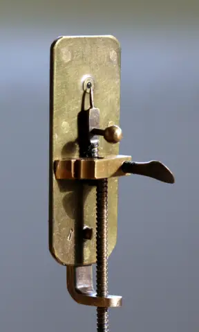
In the mid 1670s, Leeuwenhoek, a Dutch scientist and tradesman used his skills as a lens grinder to develop a microscope that was capable of higher magnification. His microscope model was very small (about two inches long and one inch across). The microscope was composed of two thin metal plates riveted together with a small bi-convex lens in between. this microscope was capable of providing magnifications of between 70 and 270x.
Leeuwenhoek's lenses were of great quality compared to others during that period with a thickness of one millimeter and curvature radial of about 0.75 millimeter. Using this microscope, it was possible to observe bacteria.
19th Century
Joseph Jackson Lister (1826)
In 1826, Joseph Jackson Lister, an English wine merchant and scientist was able to develop an achromatic lens thereby eradicating the chromatic effect (spherical aberration). Here, Lister used several weak lenses together at given distances resulting in great magnification without blurring images. This was a great breakthrough in microscopy and helped make microscopes important tools in medical research.
* In 1874, Ernst Abbe came up with the theoretical resolution of a light microscope. Here, Abbe had developed a formulae correlating resolving power to the wavelength of light making it possible to calculate the theoretical maximum resolution of a microscope.
20th Century
The 20th Century saw further developments in the field of microscopy, producing various microscopy techniques that have become very important today. These include:
The Transmission Electron Microscope (1931) - This was designed and built by Ernst Ruska and Max Knoll from the ideas of Leo Szilard. This microscope used electrons instead of light.
Phase Contrast Microscope (1932) - The phase contrast microscope was developed by Frits Zernike in 1932 for imaging transparent specimen. Imaging of samples using this microscope without using stains is achieved by using interference instead of absorption of light.
Scanning Electron Microscope (1942) - This was developed by Ernst Ruska and worked by transmitting electrons across the surface of the sample.
Confocal imaging principle (1957) - This technique was developed by Marvin Minsky and uses the scanning point of light to provide a slightly better resolution compared to microscopes that use light. Through this technique, it is easier to observe virtual slices through thick specimen.
First CAT scanner (1972) - This technology was developed by Godfrey Hounsfield and Allan Cormack and involves the combination of many X-ray images (with the help of a computer) for the purposes of generating cross-sectional views and 3D images.
Electron backscatter patterns observed (1973) - Developed by John Venables and CJ Harland in 1973, this technology is used to provide quantitative microstructural information about such samples as metal, minerals and ceramics among others.
Confocal laser scanning microscope (1978) - Developed by Thomas and Christoph Cremer, the confocal laser scanning microscope helps scan objects using a focused laser beam.
Scanning Tunneling Microscope (1981) - In 1981, Gerd Binnig and Heinrich Rohrer developed this technique that is used for measuring atom interactions.
Green Fluorescent Protein (1992) - While Green fluorescent protein was discovered in 1962 by Osamu Shimomura, Frank Johnson and Yo Saiga, it was cloned in 1992 with its derivatives being used in fluorescence microscopy.
Now take a Look at these Microscopy Techniques of Modern Day!
Return from Microscope Timeline to History of the Microscope
Return to MicroscopeMaster Home
Find out how to advertise on MicroscopeMaster!


