Lymphocytes and Microscopy
Staining, Observations, Discussion
Lymphocytes are leukocytes that develop from the common lymphoid progenitor. Although they primarily reside in the lymph nodes, they increase in size and increasingly divide once activated and migrate to the infected tissue where they destroy the infecting pathogen.
The different types are differentiated by their respective cell surface receptors and specific function with regards to immunity.
They include:
- B cells (B lymphocytes)
- T cells (T lymphocytes)
- Natural killer cells
For a healthy person, lymphocytes make up between 25 and 30 percent of the total leukocyte population, which translates to between one and two trillion of these cells in a healthy individual.
Apart from their ability to quickly respond to invading pathogens, some (particularly the B and T lymphocytes) have a unique characteristic in that they can retain memory of antigens they may have previously encountered. This makes it possible for these cells to immediately destroy such antigen when they encounter them again. And so, they are also referred to as memory cells.
Lymphocyte Microscopy
To identify these cells in a blood smear, Wright's stain can be used. This is an important stain that is often recommended for differential staining of blood smears or bone marrow.
Requirements
- Blood sample immediately drawn or stored in an EDTA tube.
- Glass slides (2 or 3)
- Cover slip
- Compound microscope
- Wright's stain
- Jar/beaker
- Drying rack
Procedure
- Place a drop of blood onto a clean glass slide
- Using another clean slide or cover slip at an angle, spread the drop of blood (or bone marrow) to create a thin film of blood
- Place the slide on a rack and allow to air dry
Staining
- Once the slide is completely dry, use a pipette to add 1.0ml of Wright's stain solution on the thin film of blood and allow to stand or about 3 minutes
- Add a few drops of distilled water or phosphate buffer (pH 6.8) and allow to stand for about 5 minutes
- Rinse the slide using water
- Allow the slide to dry and wipe away any excess stain at the back of the slide
- View the slide under the microscope starting with low power
- At 1000x, use oil immersion
Observation
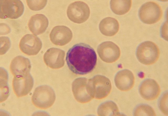
When viewed under the microscope, lymphocytes will appear dark purple with a deep bluish nucleus and a sky blue cytoplasm.
Discussion
Like monocytes, lymphocytes are agranulocytes, which simply means that they do not have granules. However, some of the larger cells (7-10um) may contain a small amount of scattered granules.
Ranging between 20 and 30 percent, they are the second largest population of leukocytes, which makes it easier to identify in a blood smear. While both the B and T cells are produced in the bone marrow, T cells go on to mature in the Thymus.
B Cells, T Cells and Natural Killer Cells
As already mentioned, their primary function is to fight and destroy antigens. This task is achieved through two major types of responses namely; humoral immunity and cell mediated immunity.
Whereas humoral immunity relies on the capacity to identify antigens to be destroyed, cell mediated immunity involves the active destruction of both the infected cells and cancerous cells.
In the presence of antigens, B cells are activated, which results in the creation of antibodies specific to the antigen in question. As such, they play an important role in humoral immunity.
Given that the antibodies remain in circulation, they are able to identify the specific antigens in the future (memory cells).
On the other hand, T cells play an important in cell mediated immunity. Here, cells move to the site of infection where they attack and destroy the foreign particles.
There are different types of T cells including:
Cytotoxic T cells - Responsible for destroying cancerous cells or infected cells.
T helper cells - Participate in the production of antibodies in addition to the activation of given T cells.
Regulatory T cells - Regulatory T cells are also known as suppressor cells and play an important role of suppressing given responses by both B and T cells.
* Natural killer cells play an important role in the rejection of tumors as well as infected cells. However, their response tends to be non-specific compared to T and B cells.
Return to Agranulocytes main page
See Also: Basophils, Neutrophils, Monocytes, Eosinophils
Return to White Blood Cells Main Page
Check out our page on Blood Smear and Cell Staining.
Return from Lymphocytes to MicroscopeMaster Home
Find out how to advertise on MicroscopeMaster!


