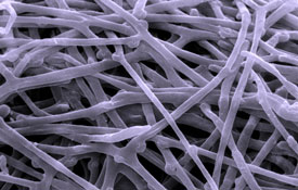
Hyphae
Production, Structure, Morphology, Types
Essentially, hyphae (singular; hypha) are the long, tubular branching structures produced by fungi. However, they can also be found in a number of other organisms such as oomycetes.
Hyphae in fungi vary in structure and serve different functions from one species to another.
Some of the most common hyphae include:
- Septate hyphae
- Coenocytic hyphae (Non-Septate hyphae)
- Pseudohyphae
* Unicellular fungi like yeast do not necessarily produce hyphae
Production
To understand how hyphae are formed in fungi, it's important to understand the life cycle of fungi. The life cycle of fungi starts with the production of spores, which are produced in the fruiting bodies of the organism.
Once the spores are released/dispersed into the surrounding environment (by wind, animals etc), they start to germinate to produce hyphae, which then develops further to form the mycelium. However, this is also dependent on the type of environment the spore lands on.
With sufficient nutrients, warmth and moisture, spores are able to germinate better compared to environments without such suitable/favorable conditions.
Growth of these fungus, and thus the hyphae, occurs at the tip of these tubular structures. In this case, new cells are continually formed at the tip of the hyphae as the structure elongates further. This occurs through a process referred to as apical elongation.
* Some hyphae are divided into cells by a septa while others are not divided.
Hyphae Structure and Morphology
Hyphae, as mentioned, grow from the spore/germ. Here, the first hyphae cell is produced and continues growing out at the apex. While some of these tubular structures can be seen with the naked eye (in large numbers) an individual hypha is a microscopic tube like structures that contain a cytoplasm (multinucleate cytoplasm) that is surrounded by a plasma membrane.
In addition to the plasma membrane/cell membrane, hyphae is also surrounded by a tougher (compared to the plasma membrane) cell wall. The cell wall is made up of chitin, which is itself a long chain of N-acetylglucosamine (polysaccharide that contains nitrogen).
One of the biggest advantages of the chitin cell wall is that it is both strong and flexible. This allows the hyphae to continue growing and elongating in any direction. This, however, is aided by the lysis of the cell wall at the tip of the hyphae.
During elongation, where cells are added to the tip of the hyphae, the cell wall undergoes lysis (degradation) allowing for cells to be added at the apex for hyphal elongation. At the same time, new cell wall is created to protect the new cells as the hyphae continue growing.
Mechanism of Hyphal Growth: Apical Growth
In fungi, hyphal growth is made possible by a number of organelles.
These include:
- Actin
- Microtubules
- Vesicles
- Golgi body/apparatus
- Spitzenkörper
Essentially, the Spitzenkörper, which is thought to be produced from the Golgi bodies, has been shown to play an important role of organizing/regulating hyphal growth. For this reason, they are found near the hyphal tip during epical elongation.
During this process (apical elongation) the cell's vesicles move towards the tip of the hyphae (towards the plasma membrane) where they release various enzymes and other compounds.
The enzymes released by these vesicles are involved in the lysis and synthesis of the cell wall. Following lysis of the wall, cell division and growth allows for the hyphae to be elongated at the tip.
Once the new cell has formed, the enzymes and other compounds at the tip start synthesizing a new cell wall around the new cell to protect it. This is then followed by the strengthening of the actin cap.
Types of Hyphae
Generally, fungal hyphae are divided into septate and non-septate hyphae.
Septate Hyphae
Septate hyphae are termed septate because they form structures known as septa between the cells. Unlike the non-septate hyphae, the septate hyphae, found in organisms such as Aspergillus species, divide the hyphae into several cells along the hyphae thread.
While the hyphae are highly divided, the septa have pores that allow for various material to pass through from one cell to another. Apart from these material and nutrients, the septa also allow for such organelles as ribosome to move from one cell to another. This is particularly important for newly formed cells at the hyphal tip.
* Apart from simply compartmentalizing cytoplasm in different cells on the hyphae, septa also plays an important role of preventing the flow of nutrients and some organelles when some of the cells in the hyphae die. This is made possible by septal plugs (Woronin bodies) that block the pores in the septa.
* On the other hand, septa (transverse septa) play an important role of dividing or separating the reproductive cells of the fungi from the hyphal threads. This is particularly important when some parts of the elongated hyphae are damaged.
There are two different of septa.
These include:
- Primary septa- formed during nuclear division and tend to separate the nuclei formed from the parent nucleus.
- Adventitious septa- The adventitious septa are not formed during nuclear division. They have also been shown to be particularly prevalent when there are changes in the concentration of the protoplasm.
Non-Septate Hyphae
Also referred to as aseptate hyphae, the non-septate lack septa and therefore tend to be elongated cells without any divisions. Because they are made up of individual, elongated cells, non-septate hyphae tend to be shorter and more erect.
They are also less branched when compared to septate hyphae. Some of the organisms that have non-septate hyphae include Mucor and zygomycetesamong others.
Hyphae may also be described according to their general appearance.
The following are types of hyphae based on morphology:
- Plectenchyma- Hyphae that appears to be fused together in a bundle
- Rhizomorph- Rhizomorph resemble branched cords and are used to obtain nutrients
- Appressorium- Appressorium are a type of hyphae cells that some fungi use to infect various plants
Types of hyphae based on their cell wall:
- Generative hyphae- Generative hyphae are characterized by a thin cell wall and greater number of septa
- Binding Hyphae- Compared to generative hyphae, binding hyphae have been shown to have a thicker cell wall and tend to be highly branched
- Skeletal hyphae- Skeletal hyphae are characterized by a long and thick cell wall, but few septa compared to generative hyphae.
Functions
For different species, hyphae are modified to serve different functions. For instance, such hypha as Appressorium, Haustoria and Mycelial strand among others play an important role of absorbing nutrients from the substrate. These types of hyphae are modified differently in a manner that allows them to access and obtain nutrients more effectively.
Some of the fungi need more specialized hyphal structures to obtain nutrients from the cells of the host they infect. For instance, the fungi Cordceps, that lives in insect hosts use haustoria (a type of hyphae) to obtain nutrients inside the insect as the hyphae grow and spread inside the host insect.
Lastly, hyphae also play an important role of producing enzymes and other substances that allow the organisms to breakdown nutrients and making absorption easier.
* Mycelia also develops from fungal hyphae
Pages to learn more about specific types of fungi:
Aspergillus - Characteristics, Types and Morphology
Alternaria - Classifications, Characteristics and Pathogenesis
Trichoderma - Classification, Characteristics and Reproduction
Pezizomycotina - Habitat, Examples, Classification and Reproduction
Claviceps - Different Species, Structure, Morphology and Life Cycle
Verticillium - Characteristics, Life Cycle, Morphology and Culture
Botrytis - Species, Habitat, Life Cycle, Infection and Culture
And the Main Page of studying Ascomycota
Return from Fungal Hyphae to Main Fungi Page
Return to MicroscopeMaster home
Resources
Charles Drechsler. (1953) Botany: Morphological features of some fungi capturing and killing amoebae.
Martin Tegelaar and Han A. B. Wosten (2017) Functional distinction of hyphal compartments. Scientific Reportsvolume 7, Article number: 6039 (2017).
Links
http://eagri.org/eagri50/PATH171/lec03.pdf
Find out how to advertise on MicroscopeMaster!

![Hyphae by AHiggins12 [CC BY-SA 3.0 (https://creativecommons.org/licenses/by-sa/3.0)], from Wikimedia Commons Hyphae by AHiggins12 [CC BY-SA 3.0 (https://creativecommons.org/licenses/by-sa/3.0)], from Wikimedia Commons](https://www.microscopemaster.com/images/512px-HYPHAE.png)


