Heterotrich
Examples, Classification and Characteristics
Overview
Belonging to the Order Heterotrichida, class Heterotrichs consists of a variety of free-living (free-swimming) ciliates commonly found in marine and freshwater environments as well as benthic (lowest level in lakes, oceans etc) and planktonic habitats.
As compared to some of the other ciliates, class Heterotrich is composed of some of the largest and colorful species. As free-living organisms, heterotrichs feed on a range of small organisms and other food sources present in their environment.
Some of the most common members of this class include:
- Folliculinids
- Peritromus
- Gruberi
- Protocruzia
- Condylostoma
Classification of Heterotrichs
· Infrakingdom: Alveolata - A major group belonging to the domain Eukarya
· Phylum: Ciliophora - Protozoa characterized by the presence of cilia on their body surface
· Class: Heterotrichea
Characteristics
Reproduction
As is the case with many other ciliates, asexual reproduction through binary fission is the main mode of cell division. However, under certain circumstances, they reproduce sexually through conjugation.
During favorable environmental conditions, Heterotrichs reproduce and increase in numbers through binary fission. Here, the process starts with the division of the nucleus through mitosis followed by the division of the cytoplasm.
Ultimately, the cell constricts in the middle allowing the two new daughter cells to separate. This results in the production of two identical daughter cells with identical DNA. Depending on the species, the entire process may take about 3 hours from start to end.
During sexual reproduction (conjugation), some of the species have been shown to release chemicals that influence compatible mating partners to prepare for the process. However, for the most part, this mode of reproduction is influenced by unfavorable environmental conditions.
Here, the process starts with two compatible cells lining up next to each other (parallel to each other). Once the cells line up, the nucleus undergoes a series of nuclear (micronuclei) division.
Here, the nuclei also undergo meiosis which results in the production of haploid micronuclei in each of the cells. These nuclei then disintegrate with only a single micronuclei remaining in each cell. Then there is division through mitosis to produce a pair of haploid nuclei referred to as gametes.
As nuclei division ends, a channel starts forming between the two cells which allows them to exchange one gamete each. These gametes fuse and undergo further mitotic division to produce a total of four micronuclei in each cell.
Whereas two of these nuclei form the micronuclei, the other two form the macronucleus of each cell. As compared to binary fission, this form of reproduction does not increase the number of the organism. Rather, it allows the cells to exchange genetic material.
* During reproduction, the nuclei are divided by extramacronuclear microtubules.
Morphological characteristics of Heterotrichs
As previously mentioned, this class contains some of the largest ciliates in nature. For instance, Spirostomum species have been shown to grow to lengths of about 4,000um (4mm). As such, they are large enough to be viewed with the naked eye. Apart from being large in size, some of the species are colorful, ranging from blue to pink in color.
Folliculinids, some of the most common ciliates, may appear blue or green-blue when viewed under the microscope. Using their vase-shaped loricae, they are able to remain attached to the substrate (adult forms of these organisms are sessile).
Some of the species are ubiquitous in their environment and use cilia to swim from one location to another (A majority of the species are free-swimming).
* The colorful nature of heterotrichs is the result of pigmentocysts (cortical vesicles).
* Pigmentation among some of the species is suggested to deter their predators.
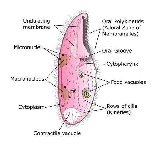
Apart from size and coloration, heterotrichs are also characterized by a conspicuous adoral zone of the oral polykinetids. Their oral cavity is surrounded by cilia structures that are significantly different somatic cilia (which is shorter).
For this reason, they are among ciliates with dikinetid cilia. Their bodies (cell body) are also contractile and can either elongate or shorten. Whereas elongation of the body is made possible by postciliodesmal microtubules, they are shortened by contractile myonemes.
In all species, the feeding membranelles (supported by oral polykinetids) are arranged in a collar and lapel manner over the anterior part of the cell. This arrangement ends up at the mouth part.
When viewed under the microscope, it can be seen beating in a wave pattern. This not only draws water towards the cell, but also various food particles that are trapped and ingested.
Some of the most common prey of heretotrichs includes:
Return from Heterotrich to MicroscopeMaster home
References
D. J. Patterson and M. A. Burford. (2001). Guide to Protozoa of Marine Aquaculture Ponds.
Hartmut Bick. (1972). Ciliated Protozoa.
Lynn, D. (2008). The Ciliated Protozoa.
Links
https://link.springer.com/chapter/10.1007/978-1-4020-8239-9_6
https://www.ncbi.nlm.nih.gov/pubmed/17880503
Find out how to advertise on MicroscopeMaster!


