- Forensic Pathology -
Microscopy Analysis and Histopathology
|
Forensic pathology is an area quite different from the other subdivisions of scientific study, as it focuses on the numerous causes of death as opposed to illnesses and their cures or treatments. The field encompasses some typical aspects of medicine, such as collecting the medical history of the deceased, obtaining samples and toxicology, as well as blood and DNA analysis. |
These techniques offer a quick and thorough analysis that supplies the initial hypothesis as to the possible cause of death, thus laying the foundation and providing direction for the remainder of the investigation.
The differences between forensic pathology and other fields of scientific study is that it also investigates deaths not caused by diseases but rather, people. Murders caused by firearms, knives, asphyxiation, drowning or blunt force trauma, along with car accidents and rapes, are all covered by a forensic pathologists autopsy.
Additionally, this specialty is also responsible for reviewing and interpreting crime scenes, along with their reports and witness statements, so that one can use deductive reasoning and logic to assist in determining the cause of death.
Histopathology
Histology is the study of human tissues, from their complex composition to their many purposes in the body.
This study is particularly important in forensic pathology because the tissue reacts to pathogens, temperature, oxygen deficiency, chemical imbalance and damage in various ways, which can indicate a cause of death but is seen as a super-specialised branch after pathology.
Each internal and external element is responsible for a specific reaction in the tissue’s cells, many of which a medical examiner may see by staining, testing and closely examining samples.
This information helps to deduce not only the cause of death but also offers an approximate time and chronological order of events. For instance, histology can tell the examiner if the deceased drowned and then suffered bruises or if a rape occurred prior to asphyxiation.
This may seem trivial to an average person but it is quite significant in forensic pathology because the medical examiner needs to pass this information on to those responsible for the criminal investigation and may also need to testify in court.
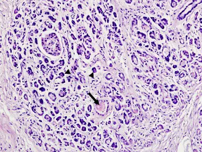
In order to study one or several areas of body tissue in the deceased, a forensic pathologist must remove an extremely thin section of the tissue as part of the autopsy. The medical examiner then processes the tissue sample by mounting it onto a glass slide and staining it so that the different cellular structures are noticeable, making examination via traditional light, scanning electron or other microscope thorough and simple.
Forensic histopathology is very useful in the case of a sudden death where trauma and poisoning need to be ruled out. As well, it plays an important role when a trivial trauma worsened or augmented the progression of disease. Forensic pathologists look at histopathology only in required cases and not routinely especially with an increased workload.
The medical examiner may also cut other tiny sections of tissue for additional tests, such as those involving freezing, specialty stains and immunohistochemistry, all of which may contribute new information to the scientific community and help find cures or treatments.
Once the results of the tissue analysis are available, which should provide answers by eliminating some possibilities while confirming others, the forensic pathologist can come to a conclusion as to the individual’s cause of death.
Interesting Read: Histology Slides - For Hobbyists, Academic Institutions and Research Labs.
Forensic Pathology and Microscopy
A traditional light microscope serves many purposes in a variety of fields but its range of abilities and the materials studied are always the same, though that does not diminish the importance of this device in scientific research.
The light microscope provides direction in forensic pathology by giving the medical examiner insight to basic organelles, ducts and molecules. The forensic pathologist can use the microscope to see the types and numbers of cells present, as well as their anatomical arrangement.
To obtain a better image of a sample, a forensic pathologist may use a light microscope with more advanced features, such as cell imaging, fluorescence or one that comes with software so that the picture may be visible digitally on a computer screen.
Some light microscopes automatically adjust the light, have a motorized stage or one that scans the material, all of which add to the quality of the sample image.
One of the most important types of light microscopes in forensic pathology are those with cameras, as they help to preserve the images if the tissue, which are often evidence in crime investigations and trials.
The right model can provide clear images with the exact colors of the sample under review, a feature that helps to easily identify the normal from the abnormal.
Comparison Microscope
A comparison microscope is used in forensics enabling the experts to literally compare and analyse two specimens side by side. It consists of two separate light microscopes attached by an optical bridge. The two specimens are viewed concurrently in a split-view window.
Scanning Electron Microscope Analysis
The scanning electron microscope is like a traditional light microscope on steroids because it can see material at a sub-micron level. This device creates two scanned images, both of which appear grey, though one shows the surface of the material while the other reveals the composition by making the heavier elements brighter.
The combination of superior image quality, contrast, high magnification and revelation of elements is particularly useful in forensic pathology when the cause of death is unnatural.
Traces of a bullet, knife, tool, car or other items made of some type of metal that can kill a person or participate in an accidental death are easier to identify under the scanning electron microscope because of the contrast.
Additionally, this microscope is also responsible for detecting gunshot residue from a sample collected from a suspect, a feature that can help put a criminal in jail but without it, that criminal can go free.
This type of microscope works with software that can automatically detect and classify not only gunshot residue but it is also customizable so that a forensic pathologist may use it to find other materials.
For instance, this microscope can detect traces of minerals in soil, which can contribute to or cause death if they are toxic or consumed in high quantities. It also has the potential to find very small pieces of bone that might remain after a fire.
Forensic pathology is an interesting, exciting and constantly changing field that requires medical examiners to stay on top of the latest information in science, ballistics and crime scenes.
This area of study not only helps to keep people safe by obtaining evidence necessary to lock up criminals but it also contributes to scientific discovery, which, when further explored, may increase the life span of human beings and help to cure or better treat life-altering illnesses.
Of further interest in Pathology:
Return from Forensic Pathology to Microscopy Applications
Return to Microscopy Research Home
Find out how to advertise on MicroscopeMaster!


