Epithelial Cells
** Definition, Function, in Urine/Sputum
Definition: What are Epithelial Cells?
Epithelial cells, found at various body sites (where they line the surfaces and cavities of body tissues and organs), are specialized components with a variety of functions. For instance, because of where they are located, epithelial cells are well suited to protect deeper tissue from the external environment (and thus from microorganisms and other factors that can cause harm).
In addition, they serve secretory and supportive functions that contribute to normal organ/tissue functions in the body. As compared to some of the other cells in the body, epithelial cells are typically characterized by specialized contacts to each other giving them a continuous sheet appearance (this sheet of cells is known as the epithelial tissue).
While they share this and a few other characteristics, epithelial cells vary in shape, size, and general appearance.
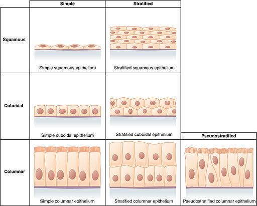
As such, they are divided into three main groups that include:
- Squamous epithelial cells
- Cuboidal epithelial cells
- Columnar epithelial cells
Origin of Epithelial Cells
In the body, different types of epithelial cells cover different surfaces in addition to being the primary components of glandular tissue.
Whereas the skin, urinary and digestive tract, etc are covered by a layer of epithelial cells known as the epithelium, body cavities like blood vessels that are not exposed to the exterior of the body are lined by a layer of cells known as endothelium (a type of epithelium covering the surface various organs/tissue located deeper in the body).
These epithelial cells originate from the embryonic layers.
Epidermis and Stratified Epithelia
Whereas the epidermis refers to the epithelial lining the skin, stratified epithelia consists of epithelial cells that line the surface of the mouth and nose as well as the anus among several other areas of the body.
These cells originate from the surface ectoderm of the external germ layer (ectoderm). These cells also give rise to a number of modified epidermal tissue including hair and nails as well as various mucous glands among others.
* Such ectodermal appendages as oral and skin glands as well as hair etc. originate from the association between the mesenchyme and epithelium.
Epithelial Cells line parts of Digestive System and Airways
Unlike epithelial cells that make up the epidermis (of the skin), cells lining the airways (respiratory tube) as well as parts of the digestive tract develop from the endoderm (innermost layer of the primary germ layer).
Here, however, it's worth noting that in the body, endodermal cells only give rise to the lining of the digestive tube. In turn, cells lining the respiratory tube as well as the lungs originate from the digestive tube.
* Apart from the lining of the respiratory and digestive tubes, some of the other structures that originate from the endoderm include glands of the gastrointestinal tract, liver as well as renal bladder.
The Lining of the Cardiovascular and Lymphatic Vessels (Endothelium)
Epithelial cells lining the vessels of the cardiovascular and lymphatic vessels originate from the middle layer of the primary germ layers known as the mesoderm. Apart from these vessels, the layer is also the point of origin for a number of epithelial cells including those lining the uterus, vagina as well as mucosa of the bladder.
Structure and Characteristics of Epithelial Cells
Types of Epithelial Cells
As mentioned, epithelial cells vary in shape and size. For this reason, epithelial cells in the body are classified in accordance to their general appearance (including the number of cell layers forming the tissue).
Squamous Epithelial Cells
Squamous epithelial cells are thin and flattened in shape. As such, they appear wider than they are tall. These cells normally line such body cavities as the abdominal cavity.
Depending on where they are found, squamous epithelial cells may either form an epithelial tissue consisting of a single layer of cells (simple squamous epithelium) or several layers of cells (stratified squamous epithelium).
The endothelium, consisting of epithelial cells that line the vessels of the lymphatic system, is made up of a single layer of squamous cells. As a result, it is thin enough to allow various chemicals to pass through.
A stratified squamous epithelium consists of several layers of squamous cells stacked on top of other cells. In this arrangement, the basal layer often consists of columnar/cuboidal cells while the apical cells (those located further from the basal surface/top layer of cells) are squamous.
A good example of stratified squamous epithelium is the skin. Here, epical cells are flattened and tend to be keratinized.
See Cheek cells under the Microscope
Columnar Epithelium
As compared to squamous cells, columnar are taller than they are wide. As a result, they appear elongated (column) when viewed under the microscope. The nucleus, which also appears to be elongated, is located closer to the base of the cells allowing various cellular activities to occur at the upper surface of the cell.
Like squamous cells, columnar epithelial cells may either form a simple or stratified columnar epithelium. Consisting of a single layer of epithelial cells, a simple columnar epithelium can be found lining parts of the digestive tract, respiratory tract as well as fallopian tubes.
The stratified columnar epithelium is composed of several layers of epithelial cells (columnar cells). This organization of cells can be found in the urethra as well as some gland ducts.
Cuboidal Epithelium
As the name suggests, a cuboidal epithelium consists of box-like epithelial cells (cube-like). These cells have a rounded nucleus that is typically located near the central part of the cell. The simple cuboidal epithelium can be found lining the surface of kidney tubules as well as gland ducts.
Stratified cuboidal epithelium is made up of several layers of cuboidal cells. These cells can be found lining the surface of the vagina, mouth as well as the esophagus.
Transitional Epithelium
Apart from the three types of cells mentioned above, there is another type of epithelial cell that forms stratified epithelium. They are known as stratified epithelial cells and can be found lining the surface of such body cavities as the bladder.
As compared to the other epithelial cells, stratified epithelial cells can change in shape allowing for expansion and contraction of organs.
Cell Junctions
Apart from the different shapes, epithelial cells are also characterized by close connections. As such, they are not separated from each other by various extracellular materials that are normally observed in other types of cells.
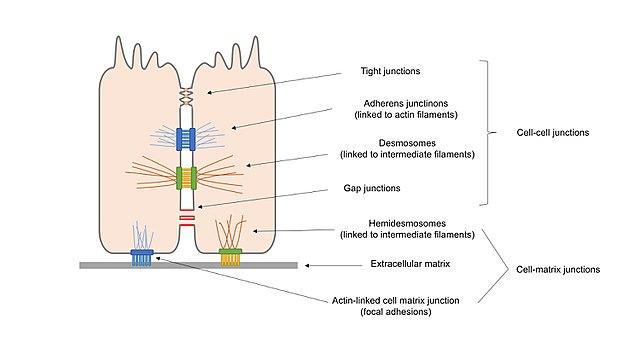
These interactions between epithelial cells are made possible by several types of connections that include:
· Tight junctions - Found between epithelial cells, where they separate them into apical and basal layers/compartments, tight junctions serve to prevent the leakage/diffusion of molecules between these layers.
It's made up of a number of molecules/proteins such as claudins that not only minimize the movement of ions or molecules between the cells, but also prevent the movement of various proteins between the cell layers.
· Gap junctions - As compared to tight junctions that create barriers between different layers of epithelial cells, gap junctions serve to allow for passage of small ions and molecules. As such, they can be described as a type of intercellular channel located between adjacent cells.
Made up of proteins known as connexins, the gap junctions are cylindrical in shape which makes it possible for ions to pass through. By allowing the passage of ions, these gaps also contribute to the changes in membrane potential from between the cells.
· Anchoring junctions - Anchoring junctions are primarily involved in stabilizing epithelial tissues.
There are several types of anchoring junctions that include:
o Desmosomes - Provide cell anchorage by attaching to the intermediate filaments (e.g. intermediate filaments of keratin)
o Adherens - Unlike desmosomes, the adheren junction provides cell anchorage by attaching to the actin bundles of cells. Made up of catenins and cadherins, this junction serves to connect neighboring cells to each other.
o Hemidesmosomes - While this junction is similar to desmosomes, it is primarily involved in the attachment of epithelial cells to the basement membrane. In doing so, it ensures that the cells are properly anchored and remain attached to this membrane.
This is particularly important in that it contributes to the overall cohesion of epithelial cells. This is achieved by anchoring the intermediate filaments of the cells to the basal lamina.
Functions of Epithelial Cells
Protective Barrier
As previously mentioned, epithelial cells are in close contact with each other. As a result, they appear as large sheets of cells that cover various surfaces and cavities in the body. This is an important characteristic in that it creates a barrier between various organs of the body and the outside environment thus protecting them from factors that can cause harm.
For instance, the skin, which consists of stratified squamous cells, creates a barrier between the internal environments of the body from the external environment thus protecting the body from various factors (microorganisms, dirt, etc) that can cause harm/damage.
This lining is also found in the body around such organs like the digestive and reproductive system etc separating these organs from the external environment.
This role of epithelial cells is also evident in such orifices as the nostrils, mouth and ear canals among others. In the respiratory system, extensions of apical epithelial cells beat in a manner that helps in the removal of trapped particles, microorganisms, and fluids.
* Epithelial cells can then be said to provide the first line of defense against various biological, physical and chemical agents that would otherwise cause harm to the body.
Selective Transfer of Material
In their protective role, epithelial cells also play an important role of controlling the transfer of material in and out of the body. For any substance to enter or leave the body, it has to pass through a given epithelium.
For instance, some of the epithelia are specialized in a manner that allows small molecules and ions to pass through. They therefore regulate the type of material passing through and are therefore involved in various mechanisms such as changes in cell potential etc.
Secretive Functions
Apart from protective functions, some of the epithelial cells are specialized to secrete and release various chemical molecules. For instance, in the small intestine, some of the epithelial cells are involved in the production of digestive enzymes involved in good digestion.
Epithelial cells lining the respiratory tube can produce mucous that prevents various microorganisms from gaining entrance into the respiratory system.
* A majority of glands consist of various types of epithelial cells. They are responsible for the synthesis and secretion of various substances and are either classified as endocrine glands (ductless) or exocrine glands that secrete substances through a duct.
As for transitional epithelial cells, these cells are capable of changing shape, they play an important role in such organs as the bladder and ureters that tend to contract and relax. As the bladder fills with urine, these cells change to squamous cells allowing the bladder to expand and hold the urine. However, when the bladder is empty, these cells change to cuboidal apical cells allowing the bladder to get back to its original size.
Some of the other important functions of epithelial cells include:
· Absorption - Where they absorb specific material required for cell and tissue functions
· Filtration - Only allowing given material to pass through
· Sensory reception - Some of the epithelial cells contain sensors that can detect changes in the environment thus allowing for proper response
Presence of Epithelial Cells in Urine
Given that epithelial cells line the urinary tract, it's normal to find small amounts of these cells in urine. In healthy individuals, these cells can be found in small amounts during urinalysis.
In the event that too many epithelial cells are found in urine during urinalysis, then this may be indicative of such underlying health conditions as kidney disease, infection of the urinary tract (e.g. cystitis and urethritis), or liver disease among others. Here, however, the number of epithelial cells in the sample can help indicate the severity of the underlying condition.
* Important precautions have to be taken during urinalysis given that the presence of squamous epithelial cells may simply be as a result of sample contamination.
* 1 to 5 squamous epithelial cells per high power field in a sample is considered normal.
* To culture epithelial cells, a urine sample is first centrifuged before being cultured. Following the culture process, cells start proliferating within a week with the colonies displaying different morphologies.
Epithelial Cells in Sputum
As is the case with urine, it's normal to find a few epithelial cells in sputum given that epithelial cells line the surface of the respiratory tract. Under normal circumstances, Gram staining technique will reveal 10 or less epithelial cells (squamous epithelial cells) in each low power field. A high number of these cells however are indicative of underlying health conditions that may require additional tests.
* To identify epithelial cells in a culture, a sputum specimen is first cultured in the appropriate culture media. Gram stains are then used to stain the sample and identify the different types of cells present.
Apart from epithelial cells, which are an indication of mucosal contamination, the sample may also contain white cells that indicate an infection.
See also: Goblet Cells, Cheek Cells under the Microscope
Return to Cell Biology main page
Return from Epithelial Cells to MicroscopeMaster home
References
Hisao Honda. (2017). The world of epithelial sheets. The Japanese Society of Developmental Biologists.
Ian G. Macara, Richard Guyer, Graham Richardson, Yongliang Huo, and Mukhtar Ahmed. (2015). Epithelial Homeostasis.
Lea T. (2015) Epithelial Cell Models; General Introduction. In: Verhoeckx K. et al. (eds) The Impact of Food Bioactives on Health. Springer, Cham
Lucía Jiménez-Rojo, Zoraide Granchi, Daniel Graf and Thimios A. Mitsiadis. (2012). Stem cell fate determination during development and regeneration of ectodermal organs.
Links
https://www.hopkinslupus.org/lupus-tests/screening-laboratory-tests/urinalysis/
https://training.seer.cancer.gov/anatomy/cells_tissues_membranes/tissues/epithelial.html
https://opentextbc.ca/anatomyandphysiology/chapter/4-2-epithelial-tissue/
Find out how to advertise on MicroscopeMaster!


