Epidermal Cells in Plants
Definition, Function, Structure and Microscopy
Definition: What are Epidermal Cells?
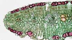
Epidermal cells include several types of cells that make up the epidermis of plants. Although they serve a number of important functions, their primary role is to protect from a variety of harmful factors (environmental stressors) including microbes, chemical compounds as well as ultraviolet light among others.
These cells are situated very close together to prevent water loss as a protective mechanism. The cell layer covers the seeds, stem, root and leaves of a plant.
In plants:
- Pavement cells
- Stomatal guard cells
- Trichomes
Structure and Functions of Epidermal Cells
Plant Epidermis
In plants, differentiation of the epidermal cells occurs during embryogenesis in a developing seed.
Like the skin epidermis, the epidermis of the plant covers the outer surface and thus covers all plant tissue from the roots to the tip. Made up of epidermal cells, the epidermis in plants also serves as a protective layer that not only prevents various microorganisms from gaining entrance into the underlying tissue of leaves and stems, but also prevents excess water loss among a few other functions.
Like the skin epidermis, epidermis of plants also consists of different types of cells that vary in morphology and serve different functions.
Plant Epidermal Cells
Pavement Cells
Pavement cells are the most common cells of the plant's epidermis. As such, they can be found covering all plant organs in any plant.
As compared to the other types of cells, pavement cells are not fully specialized. For this reason, their shapes (morphology) are not well modified for special functions as is the case with stomatal guard cells. For different plants and organs, however, studies have shown the morphology of pavement cells to vary.
For instance, in Arabidopsis thaliana, pavement cells have an irregular wavy shape that is produced during the development of leaves. In the leaves of many dicots, the shape resembles interlocking jigsaw puzzle pieces which provide some mechanical strength to the leaves.
As compared to other parts of the plant, pavement cells located in the stem and various elongated plant organs have a rectangular appearance with a long axis that is parallel to the direction of expansion (of the organ/stem). The differences in morphology have been attributed to the functions and growth forms of these organs.
* Epidermal of pavement cells in coma plants (Arabidopsis) have been shown to contain chloroplasts.
Tightly packed together, pavement cells serve to prevent excess water loss. In addition, they make up a protective layer that protects other more specialized cells located beneath.
Some of the other functions of this layer of cells include:
- Help maintain the internal temperature
- Keep the inner layers of cells in place
- Barriers to various organisms, particles and other substances from the external environment
- Separate the stomata apart (by providing tension on either side of stomata)
Stomatal Guard Cells
Stomatal guard cells are part of the epidermal tissue that serves several functions in plants.
Depending on the type of plant, the spatial arrangement of these cells is not only dependent on size, but also the shape of air-space below them. Unlike pavement cells, guard cells are more specialized with a definitive shape that allows them to carry out their functions.
With regards to structure, two guard cells form the stomata. Depending on water availability (as well as the concentration of sugars and ions), guard cells can become turgid which controls the closing and opening of the stomata pore. In turn, the closing and opening of these pores regulate gaseous exchange in and out of the leaves.
* Turgor pressure regulates the closing and opening of guard cells.
* Guard cells also contain chloroplasts that allow for photosynthesis.
Trichomes
Trichomes (epidermal hairs) are tiny hairs located on the epidermal tissue. Like stomatal guard cells, trichomes are also more specialized and thus have well-defined shapes that contribute to their functions. The trichome of Arabidopsis has been well studied and described over the years.
With large single cells measuring between 200 and 300um in length, different types of trichome have been shown to play a protective role in plants where they protect plants from predators as well as organisms that cause diseases.
Here, the trichome achieves this by either trapping or poisoning the animal to protect the plant. For some of the plants, however, trichomes simply function as barriers that protect inner tissues of leaves.
Unlike the other cells of the epidermal tissue, studies have shown that cell division is arrested in trichomes. Several rounds of endoreduplication are therefore responsible for the expansion of the cell as pavement cells continue dividing.
Cuticle
In plants leaves, epidermal cells are located on the upper and lower part of the leaf where they form the upper and lower epidermis.
The cuticle, however, is located on the upper epidermis for the most part. In plants, this is the outermost part that is secreted by the epidermis. Here, it consists of a substance known as the cutin (polymerized esters of fatty acids). On the upper epidermis, the cuticle, which is waxy in nature, acts as a water-repellent. It is also shiny and thus helps reflect off excess sunlight.
* The thickness of a cuticle in plants is largely dependent on the type of plant and where they are located.
Apart from plants, the cuticle can also be found in various organisms such as arthropods. Here, it consists of various pigments and chitin that protect the inner tissue of the organism. In human beings, however, the cuticle is the epidermis.
Epidermal Cells in Onion
Epidermal cells of onions are very simple. As a result, the epidermal tissue has become the ideal model through which students are introduced to the morphology/anatomy of plant cells.
Epidermal cells of onions also have well-defined shapes that may appear rectangular or square (or as elongated hexagonal) under the microscope.
When viewed under the microscope, it is possible to view the cell nucleus, a very thin layer of cytoplasm that can be seen in some of the cells as well as the cell walls at the boundary of each cell. These are characteristics of living cells that are capable of division and growth. Unlike epidermal cells of various plants, epidermal cells of onions have a layer of one cell in thickness.
Some of the other components of epidermal cells of onions include:
- Actin microfilaments
- A middle lamina that contains pectin
- Vacuole
Like other epidermal cells, the primary function of epidermal cells of an onion is to protect underlying tissue against such microorganisms as viruses.
See more on onion cells under the microscope.
Microscopy
Leaf Epidermal Cells
Microscopy of an onion skin is an easy and straightforward approach to observe and study epidermal cells. This is largely due to the fact that the skin can be easily prepared and viewed under the microscope. However, to observe several types of epidermal cells, then using a leaf peel is ideal.
Requirements
- Smooth plant leaf
- Compound microscope
- Microscope glass slides and coverslips
- Pair of forceps
- Water (tap or distilled water)
Procedure
- Bend the leaf to break it
- Using a pair of forceps, pull off a piece of epidermis layer from the leaf
- Place the epidermis layer on a glass slide and add a drop of water
- Place a coverslip over the sample and view under the microscope starting with low magnification
* To get a better view of the cells, slightly closing the iris diaphragm to increase contrast.
* Methylene blue stain can be used to enhance visibility.
Observation
When viewed under the microscope, stomatal guard cells are bean-shaped. In addition to guard cells, it is also possible to identify pavement cells around the guard cells.
Return to Leaf Structure under the Microscope
Return from Epidermal Cells to MicroscopeMaster home
References
Beverley J. Glover. (2000). Differentiation in Plant Epidermis Cells. Journal of Experimental Botany, Vol. 51, No. 344, pp. 497-505, March 2000.
Evaline Jacques and Kris Vissenberg. (2014). Review on shape formation in epidermal pavement cells of the Arabidopsis leaf. ResearchGate.
J. YangP. R. Verma, and G. L. Lees. (1992). The role of cuticle and epidermal cell wall in resistance of rapeseed and mustard to Rhizoctonia solani. Plant and Soil.
Miranda A. Farage, Kenneth W. Miller, and Howard I. Maibach. (2016). Textbook of Aging Skin.
Ross Carter et al. (2017). Pavement cells and the topology puzzle.
Thomas P. Colville and Joanna M. Bassert. (2001). Clinical Anatomy and Physiology for Veterinary Technicians - E-Book.
Links
https://nph.onlinelibrary.wiley.com/doi/full/10.1111/j.1469-8137.2010.03514.x
Find out how to advertise on MicroscopeMaster!

![Leaf featuring the major tissues Zephyris [CC BY-SA 3.0 (https://creativecommons.org/licenses/by-sa/3.0)] Leaf featuring the major tissues Zephyris [CC BY-SA 3.0 (https://creativecommons.org/licenses/by-sa/3.0)]](https://www.microscopemaster.com/images/512px-Leaf_Structure.svg.png)
![Opening & Closing of Stoma.As K+ levels > in guard cells,water potential of guard cells <, & water enters guard cells by Ali Zifan[CC BY-SA 4.0 (https://creativecommons.org/licenses/by-sa/4.0)] Opening & Closing of Stoma.As K+ levels > in guard cells,water potential of guard cells <, & water enters guard cells by Ali Zifan[CC BY-SA 4.0 (https://creativecommons.org/licenses/by-sa/4.0)]](https://www.microscopemaster.com/images/Opening_and_Closing_of_Stoma.svg.png)


