Does UV Light Kill Bacteria?
Ultraviolet light (UV light) is a form of electromagnetic radiation that is characterized by a shorter wavelength compared to visible light.
As part of the electromagnetic spectrum, UV light comes from the sun. However, it can also be generated from man-made sources (artificial sources of UV light) as is the case with tanning beds, lasers, and mercury vapor lighting.
Generally, UV radiation is divided into three main bands that include UVA (λ 315-400), UVB (λ280 -315nm), and UVC (λ100-280nm).
* Whereas the UV wavelength ranges from 100 to 400 nm, that of visible light ranges from 400 to 700 nm.
One of the main benefits of sunlight is that it helps the skin make vitamin D. This is especially important for normal bone development and function. However, too much exposure to sunlight, and thus to UV light, can cause damage to DNA and thus cause damage to the cells. This type of damage also occurs in bacteria and many other single-celled organisms.
* The wavelength at which ultraviolet radiation kills germ cells peaks between 260 and 265 nm (UVC λ100-280nm). This is the point at which the nucleic acids of these organisms absorb UV light. This has also been shown to be the point at which bacterial DNA absorbs UV light.
* Increase in wavelength results in reduced energy while decreased wavelength has the opposite outcome. For this reason, the UVC wavelength is more effective against bacteria and other microorganisms because it has a lower wavelength and thus higher energy.
Bacteria DNA Structure
In order to understand the impact of UV light on bacteria, it's important to understand the general structure of DNA.
Generally, the genetic material in most bacteria (DNA) exists as a single circular chromosome. However, there are some with two chromosomes while others have linear DNA.
As with many other organisms, the DNA is bacteria is a double-strand helix. The nucleotide, which is the basic building block of nucleic acids, consists of three components including the sugar, phosphate, and a nitrogenous base . The alternating sugar and phosphate make up the backbone of the structure.
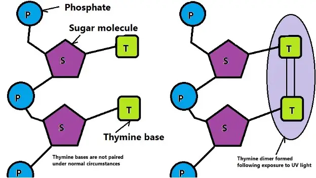
The bases, on the other hand, stick out forming the ranks of the "ladder-like" structure. Here, thymine pairs with adenine while cytosine pairs with guanine. Under normal circumstances, the bases on each strand are not linked/connected by any type of linkage. Rather, they are only paired to other bases on the other strand.
When exposed to UV light, studies have shown bases on the same strand become linked. For instance, thymine may be linked to the next thymine (through a covalent bond) on the strand to form a thymine dimer. This presents a challenge for polymerase given that it might not be able to identify the base (thymine in this case) thereby linking it with the wrong base (e.g. adenine). This results in a mutation that consequently affects other normal processes in the cell. However, this becomes a significant problem when too much mutation occurs ultimately causing the cell to die.
The following is a diagrammatic representation of a normal and mutated strand of DNA:
While the formation of thymine dimers is the primary cause of structural defects, exposure to ultraviolet radiation can also result in the formation of cytosine dimers. This has been shown to cause secondary damage to the cell.
Aside from the two dimers, UV light has also been shown to contribute to cell death through photohydration. Here, cytosine and uracil bind with water molecule elements due to the impact of UV radiation. Essentially, UV light interacts with either of the two bases causing carbon bonds to break.
In the case of cytosine, for instance, photons can break the double carbon bond resulting in the formation of a single bond between the carbons. One of the carbons then binds to hydrogen while the other binds to the hydroxyl group of the water molecule. This affects both the mechanical properties of DNA as well as its functions.
* While cross-linking of adjacent thymine is common, this type of cross-linking may occur between non-adjacent thymine following exposure to UV light. As well, cross-linking may also occur between different bases (e.g. with cytosine and guanine).
In human beings, exposure to sunlight induces the production of melanin in order to protect DNA from radiation. By shielding DNA from UV radiation, melanin prevents cell damage and potential skin cancer (sunscreen is still important to minimize the risk). The skin contains millions of cells, therefore even in the event of damage to some of the cells new ones will be produced.
In microorganisms like bacteria, however, single cells do not have this advantage. For this reason, exposure to UV radiation can kill the cell or affect cell division and cell growth.
In some cases, some bacteria have been shown to be capable of repairing the damage caused by UV light.
Two of the most common mechanisms of repair include:
- Light-dependent repair mechanism - Following exposure to UV radiation, studies have shown bacteria like E. coli to produce the enzyme photolyase (CPD-photolyase) which serves to repair the thymine-thymine dimers. The enzyme is also found in human beings. Essentially, photolyases act by reversing covalent joining created between the pyrimidines. This reverts the bases to their original monomer state thus preventing unwanted/harmful processes from taking place. In this state, cellular processes return to normal and the cells can continue growing and reproducing.
- Light independent repair - Also known as dark repair, light-independent repair does not require photoactivation to repair damage associated with UV radiation. Rather, the cell produces endonucleases which cut out and remove the damaged segments of DNA. Moreover, polymerase enzymes are produced to synthesize new DNA while ligase joins the fragments together. For instance, once the thymine-thymine dimer is detected, endonucleases cut and remove this part of the DNA . The enzyme helicase serves to unwind the DNA so that the damaged segments can be cut and removed. Once the damaged segment is removed, polymerase enzymes read the strand in order to synthesize the complementary nucleotides. This is then followed by joining the complementary pairs under the influence of the ligase enzyme.
* While some cells can repair DNA damage, excess exposure to UV light results in irreparable damage. This is because the enzymes would be incapable of keeping up or repairing such significant damage. Therefore, DNA repair often occurs when the organism has only been exposed to UV radiation for a short period of time.
In the laboratory, this is usually demonstrated by exposing bacteria (E.g. E. coli) to varying degrees of radiation. Those exposed for a short period of time survive more than those exposed for a long period of time.
Bacteriology as a field of study
Bacterial Transformation, Conjugation
How do Bacteria cause Disease?
Bacteria - Size, Shape and Arrangement - Eubacteria
How do Antibiotics kill Bacteria?
Does Salt Water kill Bacteria?
Return from "Does UV Light Kill Bacteria?" to MicroscopeMaster home
References
Goosen, N. and Moolenaar, G. F. (2008). Repair of UV damage in bacteria.
Kowalski, W. J. (2009). Ultraviolet Germicidal Irradiation Handbook: UVGI Disinfection
Theory.
Plamadeala, C. (2015). Computational Study of Cytosine Interaction With UV Light.
Links
https://www.cancer.org/cancer/cancer-causes/radiation-exposure/uv-radiation.html
https://www.medicalmate.gr/wp-content/uploads/2020/05/Ultraviolet_E_Coli_final.pdf
Find out how to advertise on MicroscopeMaster!


