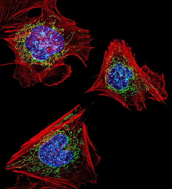
Cytoskeleton
Definition, Function, Structure and Location
Definition: What is a Cytoskeleton?
In cell biology, the cytoskeleton is a system of fibrillar structures that pervades the cytoplasm. As such, it may be described as the part of the cytoplasm that provides the internal supporting framework for a cell.
In addition to providing structural support, it's also involved in different types of movements (where it anchors various cellular structures like the flagellum) as well as the movement of cellular substances.
Composed of three components that include:
- Microfilaments
- Microtubules
- Intermediate fibers
Latest research discoveries regarding the cytoskeleton:
· The cytoplasm is involved in energy transfer as well as information processing in neurons.
· Discovery of three different nucleotides at the "barbed" end of actin filaments have helped explain why they grow much faster at one end than the other.
· Defects of the cytoskeleton can impair the immune system.
* Although the cytoskeleton provides structural support for cells, it's worth noting that it's a dynamic network of protein filaments that can be adjusted/attuned by various cues (both internal and external).
Structure and Location
Microfilaments
Microfilaments are filamentous structures of the cytoskeleton and are made up of actin monomers (f-actin). Here, globular g-actin monomers, commonly known as g-actin, polymerize to form filaments of actin polymers (f-actin). Ultimately, each strand of the filament (microfilament) is composed of two f-actin coiled in a helical fashion.
Microfilament strands have also been shown to possess positive and negative ends that contribute to the regulation of the filaments at the two ends. With regards to the development of microfilaments, studies have also found that new monomers tend to be added at the positive end at a faster rate compared to the negative end. Located at this positive end is also an ATP cap that serves to stabilize it during rapid growth.
Compared to the other components of the cytoskeleton, microfilaments are the thinnest/narrowest structures measuring between 3 and 5 nm in diameter. However, because they are made up of actin, microfilaments are quickly assembled and contribute to the proper functions of the cell.
Normally, microfilaments are located at the cell periphery where they run from the plasma membrane to the microvilli (e.g. they can be found in the pericanalicular zone where they make up the pericanalicular web/meshwork). Here, they are present in bundles that together form a three-dimensional intracellular meshwork.
* Despite being the thinnest components of the cytoskeleton, microfilaments are highly diverse and versatile.
Microtubules
Microtubules are the largest of the three components of the cytoskeleton with a diameter that ranges between 15 and 20 nm. Unlike microfilaments, microtubules are made up of a single type of globular protein known as tubulin (a protein composed of kd polypeptides and alpha and beta tubulin).
During favorable conditions, within the cell, heterodimers of tubulin assemble to form linear protofilaments. In turn, these filaments assemble to form the microtubules (hollow tube-like straws).
Like microfilaments, microtubules are also organized into bundles in cells. However, they have also been shown to be very unstable with some microtubules going through cycles of growth and shortening in their population.
During phases of shrinkage, heterodimer subunits are removed from specific ends of the tubes but added during the growth phase. The high variance of internal organization and sizes of microtubule bundles has been attributed to this dynamic instability.
* Each microtubule is composed of about 13 linear protofilaments that are arranged around a hollow core.
* Like microfilaments, microtubules are also polar structures. As such, they have two distinct ends with different charges (the positive end grows faster than the negative end).
* The dynamic instability of microtubules is as a result of polymerization and de-polymerization of the beta Bulin monomers.
In a cell, microtubules originate from the center of the cell in a hub-spoke manner. From here, they radiate throughout the cytoplasm where they serve a number of functions.
Intermediate Fibers/Intermediate Filaments (IF)
Unlike the other cytoskeleton components, intermediate filaments are made up of a large family of polypeptides. For this reason, there is a wide variety of intermediate filaments in different types of cells.
According to studies, there are over 50 different types of intermediate filaments classified into six major groups that include:
· Type 1 and II - Consists of about 15 different proteins found in most epithelial cells.
· Type III - This group includes such proteins as vimentin and desmin. They can be found in the cells of smooth muscles, white cells, and glial cells among others.
· Type IV - This group includes such proteins as neurofilament proteins and α-internexin found in nerve cells.
· Type V - An example of proteins found in this group are lamins.
· Type VI - Like nestin found in neurons.
* One of the most common proteins involved in the formation of intermediate filaments is Keratin. This is the fibrous protein commonly found in the skin and hair.
During assembly, central rod domains of two polypeptide chains are first wound around each other to form a coiled structure (dimer). The resulting dimers then come together to form tetramers that assemble on their ends (end to end) to form protofilaments. Ultimately, protofilaments assemble to form the intermediate filaments.
* Each intermediate filament is composed of about eight protofilaments.
* Unlike microtubules and microfilaments that have polar ends, intermediate filaments tend to be apolar - This is largely due to the fact that they are composed of antiparallel tetramers.
With regards to size, intermediate filaments range between 8 and 10nm in diameter- Thus the term "intermediate filaments". They are also more stable compared to the other two and thus more permanent.
Although they do not experience dynamic instability, as is the case with microtubules, proteins of intermediate filaments are often modified through phosphorylation. This plays an important role in their assembly within the cell.
In different types of cells, intermediate filaments extend from the surface of the nucleus to the cell membrane. Through the elaborate network that they form in the cytoplasm, these filaments also associate with the other components of the cytoskeleton which contributes to their functions.
Functions
Because of its localization in different types of cells, the cytoskeleton system is known for its role in providing internal scaffold that helps maintain the structural integrity of a cell.
Apart from maintaining the shape of a cell, however, it serves several other functions in cells. To get a good understanding of the cytoskeleton, it is important to look at the functions of the three components that make up the cytoskeleton.
Function of Microfilaments (Actin Filaments)
Typically, microfilaments are distributed in the motile structures of cells. They can, therefore, be found in such structures as the flagellum and cilia where they contribute to cell movement of some organisms.
Actin filaments have also been shown to be involved in the formation of such structures as the lamellipodium that allows cells to move across substrates.
Apart from cell motility, microfilaments also play an important role in the movement of various organelles. This is evident during cell division where an actin ring is involved in cell division.
Together with myosin, the filament contributes to the pinching of cells (in the middle) which eventually results in the division of cell components and consequently cell division. In the presence of ATP energy, the two have also been shown to play a role in the movement of various organelles and vesicles in a cell.
In muscle cells, actin filaments (along with myosin) are responsible for the contraction. The sliding activity of actin filaments ultimately contributes to the contraction of muscles.
* With regards to transportation of various cell components and material (vesicles and organelles etc) actin filaments act as highways or tracks through which they are transported.
Microtubules
In cells, particularly animal cells, microtubules are some of the stiffest structures with high resilience. These aspects allow them to protect cell components from various harmful forces that may otherwise cause damage.
In addition, microtubules also have the following roles/functions:
· Contribute to the architectural framework of internal cell environment - Within the cell, microtubules have been shown to help establish cell polarity by organizing cell organelles as well as other components of the cytoskeleton.
· Chromosomal segregation - Microtubules are part of the spindle apparatus that separate chromosomes during cell division. As such, they can be said to play a role in cell division.
· Transportation - Like microfilaments, microtubules also contribute to the internal transport network of a cell that allows for the trafficking of cell vesicles. In particular, this is made possible by two groups of microtubule motors, namely the kinesins and dyneins.
· Motility - Various proteins associated with microtubules help generate force and movement in such structures as flagella that contribute to cell motility.
Intermediate Filaments
For the most part, intermediate filaments serve to provide structural support for cells. In cells that experience high physical stress (muscle and epithelial cells etc), intermediate filaments help provide support that maintains the structure.
Because of their more permanent stature, as compared to other components of the cytoskeleton, intermediate filaments have also been shown to help support the cytoskeleton as a whole.
Some of the other functions of intermediate filaments include:
- Contribute to the stretching of epithelial cells
- As components of the nuclear lamina, intermediate filaments help strengthen the nuclear membrane and thus protect contents of the nucleus
- Provide support for axons as they increase in size
- They contribute to muscle contraction through the formation of bridges between Z discs
Return to Organelles main page
Return to Cell Biology main page
Return from Cytoskeleton to MicroscopeMaster home
References
A.D. Bershadsky and Iurii Markovich Vasil'ev. (1988). Cytoskeleton.
Deepa Nath. (2003). cytoskeleton. Naturevolume 422, page739 (2003).
ReHarald Herrmann and Ueli Aebi. (2016). Intermediate Filaments: Structure and Assembly.
J.E. Hesketh, and I.F. Pryme. (1996). Cytoskeleton in Specialized Tissues and in Pathological States.
Laurent Jaeken. (2007). A New List of Functions of the Cytoskeleton. Industrial Sciences and Technology, Karel de Grote-Hogeschool University College, Hoboken, Belgium.
Sabyasachi Sircar. (2008). Principles of Medical Physiology.
Links
https://www.ruf.rice.edu/~bioslabs/studies/invertebrates/microtubules.html
https://lecerveau.mcgill.ca/flash/capsules/articles_pdf/cytoskeleton.pdf
Find out how to advertise on MicroscopeMaster!

![Illustration-Proteins in prokaryotic cytoskeleton.Based on-Gitai, Z.(2005)."The New Bacterial Cell Biology: Moving Parts and Subcellular Architecture".Cell 120(5):577-586 by TimVickers[Public domain] Illustration-Proteins in prokaryotic cytoskeleton.Based on-Gitai, Z.(2005)."The New Bacterial Cell Biology: Moving Parts and Subcellular Architecture".Cell 120(5):577-586 by TimVickers[Public domain]](https://www.microscopemaster.com/images/Prokaryotic_Cytoskeleton.png)


