Copepods
Classification, Characteristics, Adaptations and Culture
Definition: What are Copepods?
|
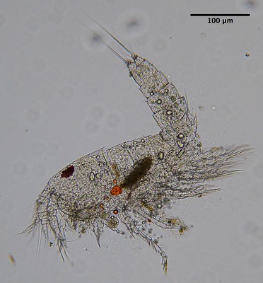
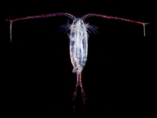
Found in natural and man-made aquatic environments, Copepods are small crustaceans that range from 0.2mm to about 20 centimeters in length depending on the species. According to studies, the subclass has the largest number of multicellular organisms compared to any other group on earth. |
Currently, over 12,500 species in the group have been described. They can be found in different aquatic environments (ranging from freshwater environments to marine environments) where they may exist as free-living organisms, symbionts, or as parasites. Depending on where they are found, Copepods are divided into planktonic and benthic forms.
* Although over 12,500 Copepods species have been identified/described to date, some researchers speculate that this only represents about 15 percent of the grand total. This, however, is hypothetical and not based on actual evidence.
Some examples of Copepods include:
- Acartia clausi
- Temora longicornis
- Calanus glacialis
- Euterpina acutifrons
- Neocalanus plumchrus
Classification of Copepods
Kingdom: Animalia - The kingdom Animalia consists of multicellular (metazoans) eukaryotes. Animals that make up this kingdom are heterotrophic and thus obtain organic sources from their surroundings.
Phylum: Arthropoda - Arthropoda is one of the largest Phyla under the kingdom Animalia. It consists of a variety of invertebrates that are characterized by a segmented body, jointed appendages, and an exoskeleton (mostly consisting of chitin).
Subphylum: Crustacea - This is a large and diverse group that consists of over 60,000 species. The majority of these species are aquatic and can be found in a variety of habitats in aquatic environments.
Members of this group are characterized by a pair of antennae, mandibles, maxillae, as well as a single compound eyes. Their bodies are also segmented into several parts with some variations between different species.
Class: Maxillopoda- Maxillopoda is a class under the phylum Arthropoda that consists of a wide variety of small animals (mostly crustaceans). With the exception of barnacles, the majority of organisms in this group feed using their maxillae. They are characterized by various head and body segments.
Subclass: Copepoda
The subclass Copepoda is further divided into the following orders:
- Calanoida
- Cyclopoida
- Harpacticoida
- Poecilostomatoida
- Cyclopoida
- Siphonostomatoida
- Branchiura
Ecology and Distribution of Copepods
Members of the subclass copepods are widely distributed in various aquatic systems across the world. They can be found in freshwater sources, estuaries, coastal lagoons, and cave environments among others. In these environments, their distribution and abundance are largely dependent on such factors as salinity, biotic and abiotic factors, temperature, and quality/quantity of food sources.
Because the majority of copepods have limited visual capabilities, they are largely dependent on chemosensory and hydro-mechanical information as they interact with their surroundings. For this reason, copepods are often used to study ecology in order to understand the manner in which such organisms interact with their surroundings and other organisms around them.
* Globally, crustaceans make up about 10 percent of the total number of invertebrates found in freshwater.
In groundwater, as well as a variety of other habitats, copepods have been shown to play a very important role in the food web. Due to their preference for certain conditions and sensitivity to anthropogenic disturbances, they are commonly used as indicators of changing environmental conditions while also providing information regarding past conditions.
Copepods are also divided into benthic and planktonic forms. The majority of free-living species, members of the order Harpacticoida, are benthic forms. As such, they are commonly found at the bottom/floor of marine and freshwater environments where they are food sources for a variety of invertebrates and fish among other organisms.
Here, they are able to use their legs and tail to swim and crawl. Planktonic forms, on the other hand, are drifters and can, therefore, be found in various water columns. Here, they are capable of swimming from point to another and feed on such organisms as dinoflagellates. However, they are also a source of food for small fish and other larger crustaceans.
Characteristics of Copepods
Life Cycle and Morphology
Generally, Copepods are small animals with the majority of species ranging from 0.2 to 5.0mm in size. Some of the species (e.g. Valdiviella) are larger and can grow to be about 28mm in length. However, studies have shown a few species, especially parasitic Copepods like Pennella balaenopterae, to grow up to 250mm in length making them some of the largest species in this group.
The general morphology of copepods varies between the different stages of development and as the organism matures. This section will, therefore, focus on the life cycle of copepods with morphological descriptions for each stage of development.
Reproduction in copepods is influenced by a number of factors including temperature and food availability etc. According to a study conducted in China, for instance, reproduction was shown to increase with a raise in temperature (up to 35°C). However, temperatures below 20°C negatively affected reproduction, significantly reducing larval development and egg hatching.
Reproduction in Copepods is through sexual means thus requiring both male and female adults. By sensing and following pheromones of the female in water, male Copepods are able to follow the trail (in aquatic environments) and find them. In the event that a male and female are compatible, they mate for fertilization to take place.
Here, the male attaches to the female's abdomen where it places a packet known as the spermatophore. This is a bag-like capsule that contains spermatozoa. Once the spermatophore is attached to the female's abdomen, sperm cells are released and enter the body of the female through the gonoduct allowing them to reach and fertilize the oocites.
Following fertilization, the fertilized eggs may be released into the water as free spawners or carried until they hatch depending on the species.
* For the most part, males are smaller than the female.
* During the mating/copulation process, the male attach to the female using their antennae.
* Following fertilization, eggs are first enclosed in the ovisac where they undergo development before they hatch.
* In some species, the eggs are covered by a tough shell that allows them to survive for a long period of time. Some eggs have been shown to survive for as long as a decade in sediments. For different species and even within the same species, studies have shown eggs to have varying characteristics with some exhibiting unique patterns or spines etc.
Hatching of these eggs gives rise to tiny larval forms known as nauplius. Early on, these forms are characterized by well-developed antennules, antennae, and mandibles at the head region. Although they do not resemble the parent at this stage, they use their appendages to swim about like their parents. During this period, many species will undergo six cycles of molting/shedding in order to produce forms that resemble the parent.
In some species, the naupliar stage produces individuals that have more than three pairs of functional appendages. These are known as metanauplius and possess bilobate maxillules and limb buds of legs.
They are also characterized by the eye morphology observed in nauplius, pigmentation, gonadal structure in some cases as well as sexual dimorphism. In some of the species, the functional maxillules in this stage are lost as the organism matures and reaches adulthood.
In the copepodid stages (free-living larvae), the thoracic and abdominal segments are distinct given that they are separated by an arthrodial membrane. While they lack naupliar endite at their antennary coxa, their post-mandibular appendages are well developed.
Some of the other characteristics associated with copepodid stages include:
- Their swimming legs are joined by an intercoxal sclerite
- The antenna loses its function
- Increase in body size as well as an increase in the number of somites
- Nine pairs of well-developed limbs at the first stage. These include antennae, antennules, mandibles, maxillules, maxillae, swimming legs, maxillipeds, and caudal rami.
In the other stages of the copepodid phase, copepods continue developing further with different structures being used for given functions. For instance, in the chalimus stage (fourth stage of the copepodid phase), parasitic species (e.g. Pandaridae, Pennellidae, and Lernaeopodidae) are able to attach themselves onto the host using their frontal filament.
Copepodid stages are followed by the pupal stages in copepods. It consists of several types of post-larval development that may include direct metamorphosis to adulthood for both the male and female sexes, indirect metamorphosis through several intermediate pupal stages in both sexes, and direct metamorphosis only in one of the sexes (with the other sex undergoing indirect metamorphosis).
This may vary from one species to another. Given that there are several stages of pupal development, each stage has certain different characteristics (morphologically).
The following are some of the main morphological characteristics in each stage:
The first pupal stage:
The body of the organism is divided into several parts that include the prosome and hirsute trunk, the antennules that consist of several vestigial setae, antennae, maxillules, as well as the maxillipeds.
Second pupal stage:
Fully developed oral appendages
Two pairs of swimming legs that resemble those of an adult female
Anatomy of Adult Copepods
The body of an adult copepod is generally divided into two regions which include the prosome and the urosome. The prosome, which makes up the forward region of the organism is divided into cephalose and metasome.
The cephalosome is the head region of the organism and consists of a single compound eye, two antennae (the first antenna is long and is used for swimming), a mandible, maxilla, and a maxilliped.
The second part of the promose, which is the metasome is divided into several segments each of which has a pair of swimming legs. The anterior region of the copepod body (urosome) is also divided into several segments with each having a specific function.
The genital segment, which is the first segment in this region is involved in reproduction where the anal segment is involved with excretion of waste material. While the urosome does not have any appendages, the last segment consists of structures known as caudal rami. It's characterized by a set of setae and is suggested to play a role in stabilization/balancing during movement.
* The body segments of copepods are also known as somites.
With regard to general anatomy, it's worth noting that the architecture varies between different species. In some of the species, the anterior body trunk consists of six broad segments/somites and a narrow posterior consisting of five somites (in gymnopleans) while others have a broad anterior consisting of five segments/somites and a narrow posterior that consists of six somites (the majority of podopleans).
For thaumatopsylloids, the anterior part of the body consists of four broad somites and seven narrow somites at the anterior part. Therefore, the general body morphology can be used for identification purposes.
Free-living vs. Parasitic Copepods
Copepods may exist as free-living forms or as parasites. The order Harpacticoida holds the majority of free-living forms that feed on various organic material (through grazing) in their environment or on given prey (predators).
In free-living copepods, the mouthparts are modified in for biting, filtering, and scraping thus making the feeding process easier. For filter feeders, the front appendages are equipped with setae that are used to create a current that directs material to the mouthparts (maxillipeds).
These materials are then filtered in the filter chamber so as to retain food material. Using modified mouthparts, they are also able to bite and seize prey. However, they have been shown to be very selective which allows them to avoid eating any material/prey with toxic substances.
The parasitic forms depend on a host (fish, whales, etc) for their nutrition. Using modified mouthpart structures, they attach themselves to given hosts and feed on their tissues. For the most part, they do not cause significant harm to the host.
This is possible in cases where the hosts are scarce. Here, a single host may be infested with numerous parasitic forms that can cause significant damage as they feed on its body tissues.
Adaptations
Copepods have various adaptations that allow them to survive and thrive in their respective habitats. Given that they have poor visibility, mechanoreception is one of the main adaptations that allow them to avoid predation. This involves the use of Mechanosensory setae which vary in shape depending on the species.
In the event of a disturbance in their surrounding, this information is received by the setae which allow them to respond quickly. According to studies, copepods have been shown to accelerate at speeds of over 600 body lengths per second within a few milliseconds in response to any disturbance. This allows them to avoid approaching predators.
In addition to Mechanoreception, copepods are also able to respond to their surroundings using chemosensory sensilla. These structures consist of several sensory cells. These structures are present on the antenna as well as the feeding appendages. While they may not have good visibility, they can use these structures to identify food sources.
Culture of Copepods
Copepods are able to multiply in large numbers within a short period of time. This makes them an important food source for fish and fish larvae.
Using various culture techniques makes it possible to enhance reproduction in order to feed fish.
Requirements
- Sieves - 500 micron mesh and a 200-micron mesh
- Tanks/containers - the size is largely dependent on the number of copepods to be cultured
- Filter bag
- Phytoplankton
Procedure
· Sieve the sample (zooplankton sample) using a 500-micron mesh in order to remove any fish or shrimp larvae that may be present in the sample
· Using a 200 micron mesh, sieve the sample again in order to eliminate the smaller zooplankton - This process may be repeated several times in order to retain adult forms of copepods for culture
· Chlorinate the water and then de-chlorinate
· Filter the water using a 5um filter bag
· In your tanks (to be used for culture), ensure that the tubes can supply oxygen effectively to the bottom of the culture
· Add the phytoplankton (e.g. Nannochlroposis) into the tank - Filling the tank half full is enough
· Place the starter culture in the container through standard acclimation procedures
· To control for temperature, the tank can be placed at about 60 percent shade and 12 hour light - Light intensity is also used to control the population of the phytoplankton
· Ideal culture conditions to be maintained include 30 to 35ppt (salinity), 25-29 degrees Celsius, and pH range of between 8 and 8.5.
Return to Plankton under the Microscope
Return from learning about Copepods to MicroscopeMaster home
References
Edward J Buskey, Petra H Lenz, and Daniel K Hartline. (2011). Sensory perception, neurobiology, and behavioral adaptations for predatoravoidance in planktonic copepods.
H.M. Verheye. (1996). Ecology and Morphology of Copepods.
Jan Heuschele and Erik Selander. (2014). The chemical ecology of copepods.
Mie H. Sichlau, Einar E. Nielsen, Uffe H Thygesen, and Thomas Kiørboe. (2015). Mating success and sexual selection in a pelagic copepod, Temora longicornis: Evidence from paternity analyses.
Santhosh B Santhosh, Anil, MK, Muhammed Anzeer F, and Aneesh KS. (2019). Culture Techniques of Marine Copepods.
Links
https://www.imas.utas.edu.au/zooplankton/image-key/copepoda
http://academics.smcvt.edu/dfacey/AquaticBiology/Freshwater%20Pages/Copepods.html
Find out how to advertise on MicroscopeMaster!


