Basic Principle of Confocal Microscope
Laser Scanning Applications
The confocal microscope provides image details that you cannot see using a conventional compound light microscope.
Utilizing state of the art technology and lasers that separate light waves, you can view images without blurred edges and in higher resolutions.
Fluorescence
In order to understand how a confocal microscope operates, an understanding of fluorescence is needed.
Fluorescence microscopy uses high intensity illumination (absorption and re-radiation of light) and the attachment of specific fluorescent dye molecules to the sample in order to see certain components of a material.
Staining slides with a fluorescent dye for study allows different wavelengths of light to be seen and by using different color dyes, various components of a sample can be viewed.
Fluorescence permits all parts of a sample to be studied by simply changing the color of the dye used when viewing the sample.
See immunofluorescence in microscopy also.
Mechanics and Application
Confocal microscopes use dichromatic mirrors to bounce light from the objective lens onto a second mirror and past a laser that separates the different colors of light waves.
When a fluorescent dyed sample passes through the laser, the microscope separates the unwanted light bands from those required to view a specific part of the sample.
In application, this type of microscope only allows a small section of the sample to be seen at a time, but allows many images to be taken quickly.
By collecting these images and combining them using a computer, you can construct two and three-dimensional pictures of the sample.

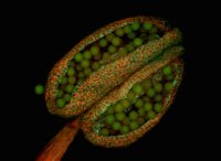
Pictured right: Anther of thale cress under confocal laser scanning microscope.
Some confocals use sound waves generated by crystals to provide faster imaging and higher resolutions.
Using this type of microscope, you may see images with 512 x 480-pixel definition.
The pinhole, the small opening where light passes through the plate and sample, of most of these microscopes, is slightly larger allowing more light bands to pass through where they can be separated easier.
Advantages
There are many advantages to using this type of microscope including the detailed study of minute objects within a sample and the ability to filter fluorescence colors providing views of a sample not visible using white light.
These microscopes provide better resolution on both the vertical and horizontal planes, in some cases up to 0.2 microns on the horizontal and 0.5 on the vertical plane.
Higher resolution, more efficient use of light and the ability to filter out unwanted light wavelengths that prohibit complete sample study are some of the reasons these specialty microscopes are highly valued in many scientific areas.
The high-speed imaging abilities of confocal instruments that provide detailed pictures were impossible until they were invented.
Optical Companies
Providing optical equipment for scientific study that is easy to operate and allows the highest confocal microscopic resolution possible is the goal of optical companies including Olympus, Leica and Nikon.
- Leica offers an easily upgraded confocal viewing system that provides detailed resolutions at the 50-nanometer range.
- The Olympus FluoFiew offers lambda scanning and unmixing of images with minimal optical image loss.
- The A1R MP microscope by Nikon utilizes galvanometer and resonant scanners that can produce images at a rate of 30 frames per second with image resolution up to 512 x 512-pixels.
All of these state of the art microscopes allow scientists to study the smallest parts of any sample and discover the intricacies of how cells and chemical bonds work.
A confocal microscope provides high definition images in two and three dimensions allowing scientists to study in detail the subtle interactions of cells, chemical reactions, and the smallest of Earth’s creatures.
For further reading, check out other microscopy imaging techiques available.
See immunohistochemistry also.
Return from Confocal Microscope to Compound Light Microscope
Return to Best Microscope Home
Find out how to advertise on MicroscopeMaster!


