Class Monogenea - An Overview
** Examples, Life Cycle, Characteristics
Overview: An Introduction to Class Monogenea
With over 4,000 species, the class Monogenea consists of parasitic flatworms that belong to the Phylum Platyhelminthes. They are commonly found in aquatic habitats where the majority of species exist as ectoparasites of fish and other organisms. A few species, however, are endoparasitic and thus invade various body organs of the host.
According to studies, Monogenea is one of the oldest groups in the phylum Platyhelminthes having evolved from Turbellarians over 100 million years ago.
While many of the species in the group share a number of morphological characteristics, such as a flattened body and being small in size, Monogeneans are divided into two major groups which include monopisthocotyleans, characterized by a hook-like structure located on their haptors, and polyopisthocotyleans which depend on a clamp-like structure for attachment.
* Monogenean was first identified by Otto Friedrich Müller, a Danish naturalist, in the 1770s. However, Muller regarded the worm as a type of leech.
* The group was named by Edouard Van Beneden, a Belgian embryologist and marine biologists - The name is derived from the Greek words "Mono" which means unique and "Genesis" which means generation.
Some examples of the Class Monogenea include:
- Polystoma integerrimum
- Ancyrocephalus chiapanensis
- Gyrodactylus salaris
- Diclidophora nezumiae
- Diplozoon paradoxum
- Dactylogyrus vastator
Classification of Monogenea
Kingdom: Animalia - Members of the Class Monogenea, which are multicellular organisms, belong to the kingdom Animalia. As such, they are eukaryotic heterotrophs and thus rely on other organisms as a source of nourishment.
Phylum: Platyhelminthes - Monogenea belong to the Phylum Platyhelminthes. They are bilaterally symmetrical and exist as free-living or parasitic organisms. They are also acoelomate with a dorsoventrally flattened body as well as a soft outer covering.
Superclass: Neodermata - Neodermata is a large group that largely consists of parasitic flatworms. These include members of the groups Cestoda, Trematoda, and Monogenea that either exists as endoparasites or as ectoparasites.
Class: Monogenea - Characteristics of this group are discussed below in detail.
Ecology/Distribution
While the majority of Monogeneans are ectoparasites, a few species are endoparasites and thus live inside the host. In various aquatic habitats, fish, and many other aquatic organisms (turtles, crustaceans, etc) act as the source of nutrition for a variety of ectoparasites that inhabit this environment.
For this reason, some researchers have suggested that over time, some monogeneans gradually became internal (endoparasites) as a response to the high pressure exerted by various ectoparasites and predators in aquatic habitats.
This is also important given that in non-fish hosts, this behavior has allowed some Monogeneans to survive in terrestrial environments. This has allowed some researchers to study coevolution through bilateral transfer.
* In terrestrial habitats, Monogeneans exist as internal (endoparasites) parasites of non-fish hosts
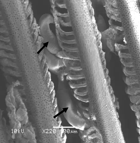
In marine, freshwater and brackish water habitats, the majority of Monogeneans can be found on the skin and gills of fish and a number of other organisms in their surroundings. They mostly exist as ectoparasites.
Here, some of the species are host-specific and can only be found attached to given hosts. However, some of the species can be found on different hosts and are therefore widely distributed.
Apart from bony fishes, Monogeneans infect such animals as rays, freshwater turtles, frogs as well as hippopotamus among others. For this reason, they can be found in aquatic habitats in different parts of the world wherever the host migrates.
They are spread from one host to another through contact (e.g. contact between different hosts) which also allows them to be spread from one habitat to another.
* Given that contact between the hosts contributes to the transmission of Monogeneans, the rate of infection escalates with an increase in contact between susceptible and infected hosts as well as with increasing host density.
Characteristics
Morphology/Structure
Like many other platyhelminthes, Monogeneans are multicellular organisms. They are very small, ranging from 0.3 to 20mm depending on the species with a leaf-shape appearance.
Some of the species are large enough ot be seen with the naked eye (Entobdella is one of the largest Monogeneans, measuring up to 2 cm/20mm). They also acoelomate and thus lack a body cavity.
For this reason, they have a flattened body (i.e. they are dorsoventrally flattened). This morphology exhibits bilateral symmetry where the right side of the body (if the organism is divided into two halves) is the mirror image of the left.
* While the majority of Monogeneans are dorsoventrally flattened, the smaller ones appear more cylindrical.
* They also tend to be colorless while others are semitransparent compared to Turbellarians which are very colorful.
Body Wall
The body wall of Monogeneans consists of the tegument that has similarities to the tegument found in trematodes. This unit (tegument) is attached to the plasma membrane both internally and externally.
In addition, microscopic studies have also revealed an outer layer of cytoplasm that connects the tegument to the parenchyma region which consists of nucleated cells. The inner membrane of the tegument is also supported by a fibrous basal lamina.
* Monogenean tegument contains secretory bodies that originate from the inner nucleated cells in the parenchyma. In addition, it may contain spike-like filamentous and microvilli structures in some of the species.
* The tegument has also been associated with absorptive activities and therefore has an important role in osmoregulation.
* In Monogeneans, the microvilli are particularly numerous in areas of the organism associated with adhesion.
The body wall of Monogeneans also consist of several layers of muscle located below the basal lamina. These include the circular musculature layer, diagonally arranged muscles as well as a thicker layer that consists of longitudinal muscles.
These layers of muscle are not striated and contain a high number of mitochondria. Here, the high number of mitochondria in these muscles is important given that they supply the energy required for the coordinated movement characterized by stretching, shortening as well as bending of the organism. This allows many Monogenean species to move rapidly in a leech-like manner.
Nervous and Sensory System
Monogeneans possess a simple nervous system that consists of three pairs of longitudinal nerve trunks that are interconnected at certain joints. These include the dorsal, ventral, and lateral trunks. The nervous system also consists of a simple brain/cerebral ganglion made up of a collection of connected nerve cells connected by commissures.
The brain is then connected to the longitudinal nerves that run to the anterior and posterior parts of the body. This allows the peripheral nervous system of the organism to run and spread in all the major organs including the pharynx, gonopore, and mouth, etc.
The nervous system contributes to a number of sensory organs in Monogeneans allowing them to effectively respond to their surroundings. The sensory system in these organisms includes photoreceptors, rheoreceptors, chemoreceptors as well as tangoreceptors.
Located on the body wall (particularly the tegument), tangoreceptors are an important part of the sensory system that allows Monogeneans to respond to pressure in their surroundings. Here, these receptors may be present in the form of ciliated pits/grooves allowing the organism to respond to different types of pressure or touch.
Chemoreceptors, on the other hand, allow the organisms to detect/sense the presence of a host (e.g. during host-host interaction) which in turn allows them to attach to the appropriate host (some Monogeneans are host-specific and will only infect given hosts).
* In Monogeneans, studies have shown the photoreceptors to be present in the eyespots/pigmented eyes or the juvenile forms (larval stage). These receptors allow the organisms to detect/sense light in their surroundings. However, some of the species may have ciliary eyes that contain a single cell.
** Monogeneans do not have a skeletal or circulatory system.
Feeding/Nutrition and Associated Structures
Ectoparasites
As already mentioned, the majority of Monogeneans are ectoparasites of fish. However, they have also been shown to be parasites of a number of other organisms including freshwater turtles.
While some of the adult forms remain permanently attached to the surface of the host, many others are browsers and move on the body surface where they feed on epithelial cells and mucus. On fish hosts, Monogenean species can be found located on the fin, gills, as well as the skin.
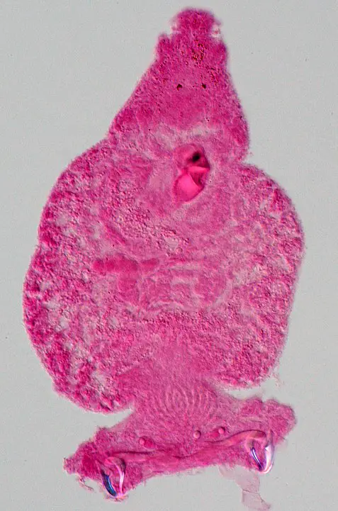
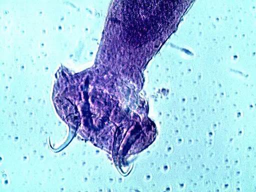
Having identified the host (using chemoreceptors), Monogeneans use a specialized organ known as a haptor (which is equipped with hooks) to attach to the epidermis of the host. While these hooks are smaller in size (marginal hooklets) in some species, they are larger in other species where they are known as hamuli/anchors.
Located at the anterior part of the body, these hooks allow the organisms to attach themselves to the epithelial tissue so that they can start feeding. Here, however, is worth noting that Monogeneans are divided into two major groups based on the structures used for attachment.
Whereas monopisthocotyleans use hook-like structures to attach to the host (these hook-like structures are located on the haptors), polyopisthocotyleans use a clamp-like structure to attach to the host.
In monopisthocotyleans, the haptor has a disc-like shape with one or several pairs of hooks. By penetrating the skin of the host, these hooks allow the organism to remain in place while they feed.
Having attached to the host using the hooks or clamps, Monogeneans also use anterior oral suckers to enhance this attachment. The mucus or epithelial cells are then sucked through the pharynx and into the intestinal caeca (sac-like/branched intestine).
For some of the species, the pharynx is glandular and produces enzymes that act on the food materials before they reach the intestine. Enzymes are also produced in the caeca to break down the food materials before the nutrients are absorbed in the gastrodermis.
* Whereas the gastrodermis in polyopisthocotyleans is adapted to absorb hemoglobin, it's adapted to absorb mucus and epithelial cells in monopisthocotylean.
* Excretion of waste material occurs through two anterolateral openings connected to the collecting ducts. They consist of flame cells and ducts and have also been associated with osmoregulation.
* Endoparasites invade various organs of the host including the urinary bladder, parts of the kidney and eyes where they feed on tissues.
* Some of the species (endoparasites) feed on the blood of the host.
Some of the signs and symptoms of infestation in fish include:
- They start swimming near the surface of the water and become lethargic
- Diminished appetite
- Their fins become clamped
- They are likely to start rubbing the bottom or sides of the tank in which they are being raised
- Loss of scale and a change in color in areas where the parasite (ectoparasite Monogeneans) are attached
- Swollen gills
Endoparasites
For endoparasites like Protopolystoma xenopodis, the adult forms infect such organs as the kidney and urinary bladder. In the case of Protopolystoma xenopodis, in particular, the adult forms can be found in the urinary bladder of Xenopus laevis (an African clawed frog). Here, the parasite produces eggs (about 9 eggs) which are then released into the external environment along with the urine.
It's worth noting that while the urinary tract provides a conducive environment (e.g. with temperatures of about 20 degrees C which promotes egg production), it also allows for the eggs produced by the parasite to be released into the environment with the urine. The eggs hatch to release oncomiracidia (in about 22 days) which are capable of actively swimming in search of a new host.
Once they come in contact with the host, they enter the body through the cloaca and gradually migrate to the kidneys for development before migrating to the urinary bladder where egg production takes place. This allows the cycle to continue once the eggs are released into the external environment.
Reproductive System and Life Cycle
All known species are hermaphrodites and thus have both male and female reproductive organs. Here, the female reproductive system has a single ovary that may either be globular, elongated or folded. The oviduct emerges from the ovary through the distal part of the ovary and runs to the ootype.
The ootype is an expanded structure that consists of melhis glands that secrete a substance that serves to lubricate the structure which promotes the movement of the eggs. The ootype is also connected to the uterus and vaginal pore through ducts.
While the vaginal structure can be seen in some species, it's absent in others. In species where it's present, it may consist of a seminal receptacle where the sperm is stored before fertilization. The seminal vesicle present in Monogeneans is itself characterized by vitellaria, which are abundant and extend to the parenchyma.
Some of the other important parts of the female reproductive system in Monogeneans include:
- Yolk glands - Consist of small follicles and are usually arranged laterally. A small duct emerges from these follicles and forms the yolk ducts
- Genital atrium
Male Reproductive System
Compared to the female reproductive system, the male reproductive system in Monogeneans has been shown to be highly variable between different species. For this reason, it can be used to differentiate between members of different species.
For instance, different species can have a few to hundreds of testes that are continuous with vessels that join to form the vas deferens. In some species, the vas deferens may have a dilation known as the seminal vesicle where sperm cells are stored. However, for other species, it simply opens up at a small papilla at the genital atrium (near the uterus).
Some of the other features of the male reproductive system in Monogeneans include:
· Complex male copulator/male copulatory organ
· Accessory piece that serves to guide the copulatory organ
· Prostate gland - present in some of the species
In mature/adult Monogeneans, the male reproductive system is the first to mature. Sperm transfer occurs through the intromission from the penile structure of the male reproductive system (of either organism). However, for some of the species, this has been shown to occur through hypodermic impregnation.
Following mutual/unilateral insemination, the eggs in the female reproductive system are fertilized and released into the environment where they develop to produce larval forms. The larval forms of Monogeneans are highly ciliated and are also known as oncomiracidium.
Apart from cilia, which allows them to swim freely, the larval forms also possess posterior hooks that allow them to be transmitted from one host to another as they develop.
Once they are attached to a suitable host, the ciliated cells are lost so that they resemble the adult forms as they mature. Therefore, the majority of Monogeneans have a more direct life cycle and do not require more than one host to complete this cycle. Once the individual matures and becomes an adult, the life cycle can continue.
* While Monogeneans are hermaphrodites, the copulation process does not always result in the eggs of both members being fertilized. On the other hand, in some cases, one individual can fertilize its own eggs (self-fertilization). However, self-fertilization is very rare.
* Once the eggs are released following the fertilization process, hatching has been shown to be influenced by such factors as shadows and chemicals.
* Depending on the species, the eggs may take between 2 and 3 days to hatch and about 10 days for the organisms to reach maturity.
See more on Flatworms:
All about Phylum Platyhelminthes
Return from Class Monogenea to MicroscopeMaster home
References
Fabiana B. Drago & Verónica Núñez. Chapter 5. Monogeneous Class.
Graham C. Kearn. (2014). Some Aspects of the Biology of Monogenean (Platyhelminth) Parasites of Marine and Freshwater Fishes.
Maxine Theunissen, Louwrens Tiedt, and Louis H. Du Preez. (2014). The morphology and attachment of Protopolystoma xenopodis (Monogenea: Polystomatidae) infecting the African clawed frog Xenopus laevis.
Patrick T K Woo. (2006). Fish Diseases and Disorders, Volume 1: Protozoan and Metazoan Infections.
Ruth Francis-Floyd, RuthEllen Klinger, Denise Petty, and Deborah Pouder. (2019). Monogenean Parasites of Fish.
Sherman S. Hendrix. (1994). Marine Flora and Fauna of the Eastern United States Platyhelminthes: Monogenea.
Links
https://encyclopediaofarkansas.net/entries/monogeneans-9034/
Find out how to advertise on MicroscopeMaster!


