Cell Wall
Definition, Function, Structure, Location, Vs. Cell Membrane
Definition: What is a Cell Wall?
Essentially, the cell wall is a complex, highly organized structure that defines the shape of a plant cell (it's also found in bacteria, fungi, algae, and archaea).
In addition to defining the shape of plant cells, a cell wall has a few other functions that include maintaining the structural integrity of a cell, acting as a line of defense against a variety of external factors as well as hosting various channels, pores and receptors that regulate various functions of a cell. As such, it's a multifunctional structure in plant cells that also contributes to plant growth.
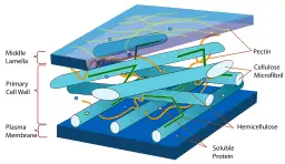
Depending on the type of plant/cells, a cell wall may contain different types of polysaccharides (carbohydrate polymers), proteins and aromatics which contribute to its multi-layered structure. For a majority of plants, this structure is divided into primary and secondary cell walls that may vary in morphology and general functions.
The three layers of a cell wall include:
- Middle lamella
- Primary cell wall
- Secondary cell wall
* Unlike plant cells, animal cells do not possess a cell wall around their plasma membrane.
Structure of the Cell Wall
The cell is part of the apoplast and thus located between the cuticle (found in some organisms) and the plasma membrane.
As already mentioned, the cell wall may consist of two or three layers depending on the type of plant of cell. These layers vary in macromolecular composition, thickness, and functions in different organisms.
While the cell wall appears as a single structure all-around a cell, it is worth noting that the structure is deposited around the cell in a series of layers. Here, the layers are deposited around the cell as the cell divides.
As new cells continue being produced around the parent cell, new layers are added around the cell with additional material being included between the plasma membrane of the cells (as well as the previously added layers).
Cell junctions between neighboring cells, therefore, play a crucial role in cell wall development given that they allow for the necessary material to be added to form the cell wall.
* The first layer of the cell wall is made from protoplasmic matters produced during nuclear and cytoplasmic division.
Middle Lamella
Located between the double cell wall of adjacent cells, the middle lamella is the first layer of the wall to be formed. Being the outermost layer (of the three layers of the cell wall) the middle lamella acts as the part that binds adjacent cells.
During cell division (particularly cytokinesis), cisternae that originate from the endoplasmic reticulum as well as pectin and phragmosomes containing vesicles are deposited at the equatorial plate. The plate becomes increasingly narrow as cells grow and increase in size. However, it's thicker at the corners.
Some of the other contents of the middle lamella include:
- Calcium ions
- Magnesium ions
Because the middle lamella is formed between plant cells as they continue dividing, it acts as the cement that holds neighboring cells together while also creating a boundary between the cells. When petals and leaves fall, the middle lamella dissolves, which allows cells to separate.
Primary Cell Wall
The primary cell wall starts forming once the cell plate (middle lamella) is complete. Deposits to this layer start before cell growth and proceed as the cells continue developing. As such, it continues expanding over the lifespan of the cells.
Some of the primary components of this layer include:
- Cellulose
- Hemicellulose
- Pectin
As the cell matures, cellulose microfibrils are formed and added into the outer part of the layer. While the outer side of the layer consists of an irregular network of these microfibrils, microfibrils are perpendicularly oriented to the cell axis.
As the cell grows, the primary has been shown to become thinner. As a result, it might prove difficult to separate this layer from the middle lamella. The two are therefore described as a single structure/layer known as compound middle lamella in some books.
Apart from cellulose, the primary cell wall also contains high amounts of lignin that has been shown to form covalent cross-links with pectin and protein in the layer. On the other hand, xyloglucan in the layer is bound to cellulose (through hydrogen bonds) and to protein and pectin through covalent bonds.
* Covalent bonds have also been identified between carbohydrates and lignin forming lignin-carbohydrate complexes.
Some of the other components of the primary cell wall include:
- Phenolic esters
- Enzymes
- Proteins
- Minerals such as boron and calcium
Apart from providing structural and mechanical support to cells, primary cell walls have been shown to play an important role in the ripening of various fruits and vegetables. This, therefore, makes it the primary textual components in most of the food-producing plants.
Here, changes in the structure and composition of the contents in the layer cause the ripening of fruits and vegetables.
Characteristics of the primary cell wall:
- Found in all plants
- Elastic in nature and ranges between 1 and 3 um in thickness
- Fibrils are loosely arranged
- Elongates over time
- In the cells of dicot plants, the primary wall consists of about 30 percent pectic polysaccharides
- Consists of between 15 and 30 percent of cellulose
- Contains about 20 percent of proteins
Secondary Cell Wall
As compared to the primary wall, the secondary cell wall is thicker and thus stronger ranging between 5 and 10um in thickness. Being a thicker layer, the secondary wall can also be divided into several layers including S1, S2, and S3.
Based on various studies, these sub-layers have been shown to vary in orientation, cellulose composition as well as the general direction. When the cells stop growing (or when they start to differentiate in some plants) they start to deposit an additional layer beneath the primary layer.
Here, a lignified secondary wall is gradually deposited into the layer and where it develops to provide mechanical strength and contribute to water transportation in a plant.
Some of the main components of the secondary layer include:
· Pectin - refers to a group of complex polysaccharides. They are all characterized by 1, 4-linked α-D-galacturonic acid. Examples of pectin include: rhamnogalacturonans, galacturonans, and Homogalacturonans.
· Cellulose - refers to an organic compound/polysaccharide that is composed of 1,4-linked β-D-glucose.
· Hemicellulose - Hemicellulose refers to a polysaccharide with a similar structure to cellulose. However, hemicellulose has a branched structure. Good examples of hemicellulose include galactomannan, glucuronoxylan, and glucomannan.
· Lignin - refers to a group of complex organic polymers found in vascular plants as supportive tissue (they can also be found in some algae).
* Composition of these components vary from one plant to another.
In the secondary wall, cellulose acts as the load-bearing unit where its microfibrils bind to the hemicellulose (e.g. xylan) in order to form the basic framework of the layer. Addition of lignin (which is hydrophobic and inert in nature) to the two compounds (cellulose and hemicellulose) contributes to the mechanical strength as well as hydrophobicity to the secondary cell wall.
While the primary wall is typically found in food/fruit-producing plants, secondary walls can be found in the cells of various plants used for structural purposes (timber, grasses, etc) or food for some animals. Here, the secondary walls exists in the form of fibers/woods and thus used in day to day life.
Some of the main characteristics of the secondary cell wall include:
- Ranges between 5 and 10um in thickness
- Has a number of pores
- Is only present in some cells
- Water content ranges between 30 and 40 percent
- Microfibrils are elongated and compact
* Secondary cell walls can be found in tracheids, sclereids and xylem fibers.
Reaction Wood Layers
Unlike the other cell wall layers, some plants will form a tough layer composed of high levels of gelatin and some cellulose as a response to various stressors. For instance, when a tree branch or stem is bent/tilted (thus likely to break) the secondary walls differentiate to form another layer consisting of additional layers of gelatin or lignin that provide the structural support required.
For the most part, this is a normal response to gravitational or mechanical stimuli in order to prevent the stem from breaking off. Here, the new layer is formed at the underside (in the secondary wall) of the affected part of the stem.
Contents of the wood cell wall (referred to as the gelatinous or G-layer) including such pectin as rhamnogalacturonan I and polysaccharides provide the necessary toughness that strengthens the affected area thus allowing the plant to continue growing naturally.
Bacteria Cell Wall
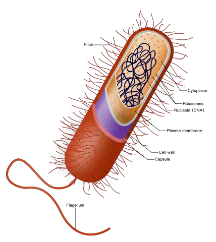
A majority of bacteria (about 90 percent) have a cell wall. However, for those without a cell wall, survival is made possible by living inside the cell of a host. Good examples of bacteria that lack a cell wall are L-form (derived from bacteria that typically have cell walls) bacteria and mycoplasma.
* Because of the bacterial cell wall, it is possible to categorize bacteria into two main groups: Gram-positive and Gram-negative bacteria. This makes it possible to identify them and develop drugs that target cell wall synthesis.
However, bacteria that lack a cell wall tend to be naturally resistant to antibiotics because they lack this structure.
Gram-Positive Vs. Gram-negative Bacteria
As with plants and a number of other organisms, the cell wall serves to maintain the general shape in bacteria (particularly for the rod-shaped bacteria) and protect the cell membrane and obtain nutrients among other functions.
One aspect that differentiates the cell wall of bacteria from that of others, however, is the peptidoglycan. This is a unique substance that has not been found in any other organism or place on earth.
In addition to peptidoglycan, some of the other components of the bacterial cell wall include:
- Lipids
- Proteins
- Teichoic acid
- Lipopolysaccharides
Components of the peptidoglycan:
· Glucose derivatives: N-acetylglucosamine (NAG) and N-acetylmuramic acid (NAM) cross-linked by tetra-peptide
· Tetrapeptide - This is a complex substance composed of 4 amino acids (D-glutamine, D-alanine, D-glutamine, and L-lysine)
Cell Wall of Gram-positive Bacteria
The cell wall of Gram positive bacteria is composed of high amounts of peptidoglycan. Here, cross-linking the tetrapeptides with a peptide interbridge produces a strong cell wall. Apart from the peptidoglycan, the cell wall of Gram-positive bacteria also contains a glycopolymer known as teichoic acid.
Some functions of this glycopolymer in the cell wall include:
- Generation of negative charge required to develop proton motive force
- Enhance rigidity of the wall
- Support cell division
- Protect the cell from extreme environmental conditions
Cell Wall of Gram-negative Bacteria
Unlike Gram-positive bacteria, Gram-negative bacteria possess a thin peptidoglycan that makes up about 8 percent of the total cell wall. This is significantly less compared to that of Gram-positive bacteria that makes up about 90 percent of the total cell wall.
While the cell wall of Gram-negative bacteria contains less amounts of peptidoglycan, their cell walls have been shown to be more complex.
Components of their cell wall include:
An outer membrane - Unlike Gram-positive bacteria, Gram-negative bacteria have an outer membrane (plasma membrane) surrounding the thin peptidoglycan layer. Although the outer membrane has many characteristics of the typical membrane, it is distinguished by the presence of lipopolysaccharide molecules.
Some of the functions of the lipopolysaccharide include:
- Contribute to the negative charge of the cell
- Protect the internal contents of the cell
- Stabilize the membrane
Lipid A - Acts as a form of endotoxin
Porins - Transmembrane proteins that form pores on the membrane
* Unlike Gram-negative bacteria, Gram-positive bacteria possess a thicker peptidoglycan layer and that retains the primary stain during Gram staining.
* In fungi, the cell wall consists of chitin and serves to provide structural support while also preventing excessive water loss.
Cell Wall vs. Cell Membrane
The cell membrane is the outer covering of all cells that encapsulates various cell organelles. It is composed of a lipid bilayer, proteins as well as some carbohydrates.
While the cell membrane can be found on covering the cells of both plants and animals, cell walls can only be found in plants and some organisms (e.g. some bacteria, etc). In these organisms, the cell wall, which is more rigid in nature, covers the cell membrane thus acting as a protective layer to both the cell membrane and contents of the cell.
* In terms of structure, the cell wall is a complex, continuous matrix mostly composed of carbohydrates and proteins while the cell membrane is made up of a lipid bilayer and has the consistency of salad oil at room temperature.
Studies have shown the cell membrane to be in direct contact with the cell wall. For the most part, this is attributed to the turgor pressure. During plasmolysis, when turgor pressure is disrupted, the plasma membrane is no longer pushed outwards and thus separates from the cell wall.
* The cell wall and plasma membrane shares a number of functions including:
- Protecting intracellular contents
- Regulating the movement of substances in and out of the cell
- Preventing excessive water loss
Unlike the cell membrane, the cell wall has the following functions:
- Strengthen and define the shape of a cell
- Provide better protection against mechanical stressors due to its rigidity as well as from various infective organisms
- Regulates expansion of the cell through its growth
- In some organisms, the cell wall acts as a food reservoir
Return to Organelles main page
Return to learning about Unicellular Organisms
Return to Eukaryotes Vs. Prokaryotes
Return to Cell Biology main page
Return from Cell Wall to MicroscopeMaster home
References
Bruce D. Kohorn. (2000). Plasma Membrane-Cell Wall Contacts. Published September 2000. DOI.
C.T. Brett and K.W. Waldron. (1996). Physiology and Biochemistry of Plant Cell Walls.
Nicholas C. Carpita, Malcolm Campbell, Mary Tierney. (2001). Plant Cell Walls.
Ruiqin Zhong, Zheng-Hua Ye. (2015). Secondary Cell Walls: Biosynthesis, Patterned Deposition and Transcriptional Regulation. Plant and Cell Physiology, Volume 56, Issue 2, February 2015, Pages 195–214.
Jasna Stevanic Srndovic. (2008). Ultrastructure Of The Primary Cell Wall Of Softwood Fibres Studied Using Dynamic Ft-Ir Spectroscopy.
Links
https://onlinesciencenotes.com/differences-between-primary-and-secondary-cell-wall-in-plants/
https://www.ncbi.nlm.nih.gov/pmc/articles/PMC2949028/
Find out how to advertise on MicroscopeMaster!

![Cell Wall or Extracellular matrix in plant- its different layer and their placement by Author: RIT RAJARSHIRIT RAJARSHI [CC BY-SA 4.0 (https://creativecommons.org/licenses/by-sa/4.0)] Cell Wall or Extracellular matrix in plant- its different layer and their placement by Author: RIT RAJARSHIRIT RAJARSHI [CC BY-SA 4.0 (https://creativecommons.org/licenses/by-sa/4.0)]](https://www.microscopemaster.com/images/Plant_Cell_Wall.png)


