Basic Components of Cell Theory
* Timeline starting from Robert Hooke *
Proposed in 1838, over 150 years after Robert Hooke’s Micrographia, cell theory is the foundation of modern biological sciences.
As microscopes became more sensitive and observational techniques allowed for the viewing of internal cellular structure, the theory expanded; but the original three tenets have remained the same.
Cellular Theory Defined
Classical cell theory, first proposed by Matthias Schleiden and Theodor Schwann, consisted of three primary points:
- All living things are made up of cells.
- Cells are the basic units of structure, function and physiology in living things.
- Living cells can come only from other pre-existing cells.
- Modern cell theory adds two additional points.
- Cells contain and pass on hereditary information during cell division.
- All cells are relatively the same in relation to chemical composition and metabolic activity.
The significance of this seemingly basic concept cannot be denied. At the foundation of almost all sciences, including biological sciences, chemistry, physiology and medicine, is cell theory.
This turning point proved on an elemental level cells are similar in the way they function and reproduce and, with the advent of more advanced observation techniques, the differences between cells relates to genetic make-up.
Robert Hooke (07/08/1635 – 03/03/1703)
Describing the appearance of a thin layer of cork tree, one of the many microscopy documentations contained in the 1665 publication Micrographia, Robert Hooke is credited with term “cells.”
Hooke likened the magnification of the cork’s box-like pores to a Monk’s living quarters, known as a cell; of Latin origin, cella is translated as “storeroom” or “small container”.

Hooke did not identify any internal cellular structure, only the external walls of the dead cork cells. Along with other scientists of that era, he thought the empty spaces might transport fluids in plant-life.
Detailed illustrations of objects observed under his crude microscope, Micrographia brought a fascination to the world of microscopy.
Hooke offered descriptions, drawings and copperplate engravings that folded out up to four times the size of the book itself.
In addition to his famous picture of a flea, illustrated observations included a fly’s eye, plant, louse as well as inorganic objects such as a needlepoint and razor blade.
Hooke was interested in microscopic and telescopic worlds, evident in his observations of the surface of the moon and astronomy related speculations posited in Micrographia.
A philosopher and architect, Hooke dedicated most of his life to science, with specific interest to optics and gravitational theory – both of which led to controversy with Isaac Newton within the Royal Society.
He was also an accomplished inventor, credited with the coil balance spring in pocket-watches, Hooke’s joint or the U-joint, found in most cars, a wheel barometer and contributions to the compound light microscope.
Contributions to the field of physics include Hooke’s law, where he explained the physical properties of linear elasticity as it related to a spring; he also made significant advances to the design of astronomical instruments such as Gregorian (reflecting) telescopes.
Hooke died long before the development of cell theory.
See more on Cork Cells under a Microscope.
Cell Theory Timeline
Cell theory can be attributed to the work of many scientists, physicians, naturalists and clergyman that took place between the mid 17th and early 19th century.
Important people and their contributions include:
- Anton Van Leeuwenhoek – saw and described bacteria in 1674; considered the first to observe single-celled organisms known as Prokaryotes
- Matthias Schleiden – German botanist; discovered all plants were comprised of cells
- Theodor Schwann – observed cells in all samples of animal tissue, therefore expanding Schleiden’s hypothesis to include animals; a German biologist and zoologist, he later discovered the sheath that encompasses nerve axons; subsequent experiments qualitatively disproved theories of spontaneous generation
- Rudolf Virchow – in 1855, published the “biogenic law”: omnis cellula e cellula, which translates into the well-known dictum “every cell stems from another cell”; he also posited all disease involved changes to the function or structure of normal cells

In 1839, Schleiden and Schwann worked together to detail the first two principles of cell theory; approximately 20 years later, Rudolf Virchow completed cell theory when he determined that cells only come from other pre-existing cells.
Although modern theory has expanded on the initial three points, the foundation established from these early findings is still relevant today.
Other notable scientists whose work validated and contributed to cell theory include:
- Francesco Redi – an Italian doctor determined that spoiled meat attracted but did not transform into flies. In 1668, this simple discovery was antecedent to the concept that cells can only come from other like-cells.
- John Needham – a naturalist and clergyman from Scotland discovered the presence of micro-organisms in soup left exposed to air; he opined a “life force” existed in all matter – organic and inorganic.
- Lazzaro Spallanzani – a biologist and abbot from Italy; preformed experiments on soup in sealed containers from 1765-67 and proved the micro-organisms that spoiled the soup were air-born – further proof that cells can only reproduce like-cells.
- Henri Dutrochet – confirmed connection between animal and plant cells; in addition, he proposed, “everything is ultimately derived from the cell”.
- Louis Pasteur – performed additional experiments on soup in the mid-1860s employing filtered air and flasks with straight necks and long S-shaped necks; he discovered air-borne microbes were able to infect the broth in regular flasks but bacteria settled at the top of the curve in the S-shaped flasks, leaving the soup unaffected. Pasteur’s work, essentially proved Vischow’s addendum.
- August Weissman – in 1880 proposed amending “biogenic law” to include living cells “can trace their ancestry back to ancient times”.
Cell Structures under the Microscope
The microscope was improved and modified for better observation of different cells and microscopic organisms. As a result, cell theory was created and modified to be what we know today.
The physiological and biological study of cells focuses on cellular structure and function.
Cells function individually, yet also come together to form organs and play a role in respiration, elimination and digestion.
At the foundation is cell theory: all living organisms are made up of cells that can only come from pre-existing like-cells – therefore, to understand behavior, genetics or disease, one must study cells.
The two primary classifications of cells are prokaryotic, which contain the domains of Eubacteria and Archaea, and eukaryotic, which encompasses protists, plants and animals.
Prokaryotes, literally translated as “before the nucleus,” contain the domains of bacteria and archaea; these organisms lack a nucleus and membrane-bound organelles, but do contain circular DNA. Unique to prokaryotes are flagellum and pilus or fimbriae.
Some prokaryotes contain organelles through infolding such as cilia, centrioles and ribosomes.
The internal and surface structure of a bacterial cells include:
- Nucleoid
- Ribosomes
- Storage Granules
- Endospore
- Capsule
- Outer Membrane
- Plasma Membrane
- Cell Wall
- Periplasmic Space
Prokaryotes replicate through binary fission.
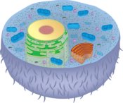
Eukaryotes are more complex organisms and, with few exceptions, contain membrane-bound organelles. Common to plants and animals cells are cell membranes, cytoplasm and a nucleus.
Organelles found in animals include:

Many animal cells also contain hair-like cilia or flagella for movement. Unique to plant cells are cell walls, plastids and a central vacuole.
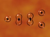
Eukaryotic cells divide via meiosis, where the cell produces gametes, or mitosis, where the cell clones itself.
What are the Differences between Meiosis and Mitosis?
With applications in almost every area of science, cell theory provides a foundation for understanding the structure and function of cells, organs and diseases.
Cytopathology is benefiting due to this foundation and, as well, pathology is advancing digitally leading to quicker and better diagnosis.
Hooke’s research was the antecedent of a theory that took numerous scientists and well over a century to devise. Schleiden and Schwann proposed the first two elements and Virchow’s “biogenic law” completed what is now the foundation of all cell-based studies.
And Amazon has not forgotten about Teachers and Educators. Useful supplies are available to aid in the teaching of microscopy and scientific concepts.
Check out the History of the Microscope here
And understanding the discovery of protists in Microscopy is important too!
Molecular Biology of the Cell 5th Edition - is a popular book in cell biology and is discussed here to further your understanding of its key features and reader feedback.
Learn more about Cell Culture, Cell Division, Cell Differentiation and Cell Staining as well as Tissue Culture Types and Techniques.
Return to Cell Biology Main Page
Return from Cell Theory to Best Microscopes Home
Find out how to advertise on MicroscopeMaster!


