Cell Membrane
Definition, Structure/Function
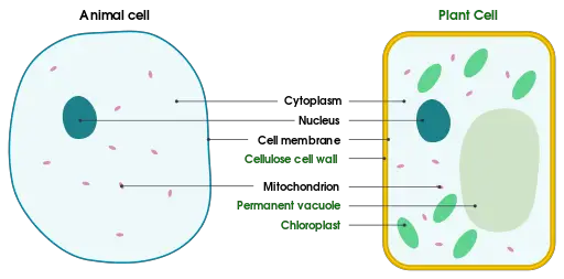
Diagram, Animal/Plant Cell
Vs Plasma Membrane
Definition
Essentially, a cell membrane is the outermost barrier that separates the internal contents of a cell in the cytoplasm from the external environment (e.g. separates the internal environment of the cell from the extracellular matrix or forms the boundary between neighboring cells). It's thin and fragile, ranging from 5 to 10nm in thickness depending on the type of cell.
In all living organisms, the cell membrane serves both morphological and functional roles varying from one type of cell to another. For instance, in amoebas, the cell membrane plays an important role in the development of pseudopodia that are involved in feeding and locomotion.
Some of the other functions of the cell membrane include:
- Transport of molecules and ions in and out of the cell
- Communication between neighboring cells
- Signal receptor
- Secretion
Structure
The cell membrane is a complex structure that consists of a phospholipid bilayer. As such, it consists of lipids in the form of phospholipids. They may also contain cholesterol and glycolipids.
Apart from these lipids, the membrane also consists of a number of other components (e.g. glycoproteins, proteins, and ions, etc) that serve a variety of functions. Here, the manner in which cell membrane components are arranged is associated with their respective functions.
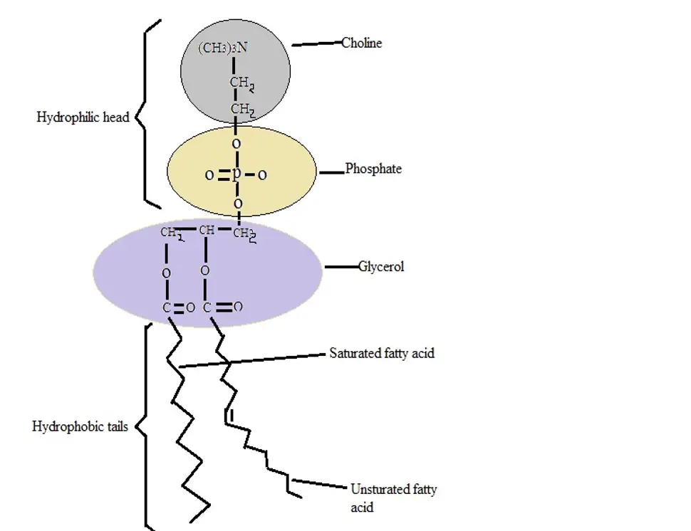
For the majority of cells, the cell membrane consists of three main components. These include glycerol, two fatty acid chains as well as a phosphate group. Therefore, for a cell membrane with glycerol as the backbone, this complex is known as glycerophospholipids (or phosphoglycerides).
Here, the glycerol, which is the backbone of the membrane, consists of three carbons, fiver hydrogen, as well as three hydroxyl groups. Attachment/linkage of the fatty acid chains (two fatty acid chains) to the glycerol occurs through a process known as esterification - the first and second carbons of the glycerol react (esterification) with the carboxyl groups of the fatty acid chains to form an ester bond.
In addition, another ester bond is formed between the hydroxyl group located at the third carbon of the glycerol and phosphoric acid. These reactions result in the production of diacylglycerol 3-phosphate (phosphatidate) which is the simplest form or precursor of the phosphoglyceride.
* The phosphoglycerides are produced through the formation of an ester bond between the phosphate group of the precursor (phosphatidate) and the hydroxyl group of given alcohols (e.g. choline, ethanolamine, and inositol among others).
While the cell membrane of eukaryotes and most bacteria consist of phospholipids, the membrane of archaeans (single-celled prokaryotes) consists of ether lipids. Here, the non-polar chains have been shown to be linked to the glycerol through ether bonds.
This type of link is particularly important for archaeans given that it's more resistant to hydrolysis. It allows these cells to survive in hostile habitats. In addition to the ether links, the membrane of these organisms consists of branched alkyl chains.
Unlike ether linkages, these chains tend to be highly resistant to oxidation and thus withstand such harsh environmental conditions as low pH, high salinity, and extreme temperatures.
* In some organisms, lipids are in the form of glycolipids (sugar-containing lipid).
Some of the other components of the plasma membrane structure include:
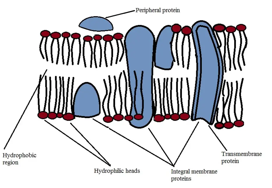
Proteins - While they are not as abundant as lipids, proteins are also an important part of the cell membrane. In the majority of cells, the cell membrane consists of two types of proteins namely, integral and peripheral proteins.
Integral membranes are embedded in the membrane (integrated into the cell membrane). While some of these proteins extend along the entire width of the membrane known as transmembrane proteins, others only extend halfway.
The proteins have one or several hydrophobic regions that anchor them to the hydrophobic part of the cell membrane in the hydrophobic tail.
Unlike integral proteins, peripheral proteins are either located inside or outside the surface of the cell membrane. They may be attached to the phospholipids of the membrane or to the integral proteins embedded in the membrane.
* The transmembrane that extends the entire width (thickness) of the membrane may consist of several hydrophobic amino acids. In this case, the amino acids are arranged in an alpha helix.
* Membrane proteins have been shown to make up about 50 percent of the total membrane mass - While they are present in smaller numbers compared to lipids, they have a large molecular mass.
Carbohydrates - Like peripheral proteins, carbohydrates are commonly found on the surface of the cell. Unlike proteins, carbohydrates are on the extracellular side of the cell membrane. Here, carbohydrates, consisting of between 2 and 60 monosaccharides, may be linked to proteins thus forming glycoproteins or bound to lipids to form glycolipids. For the majority of cells, sugars are bound to between 2 and 10 percent of lipids
Water and ions - Water is an integral component for all molecules found in living matter. As such, it can also be found in the cell membrane where it's integrated into the polar ends of the membrane (the hydrophilic ends).
Apart from water, ions have also been shown to be associated with the membranes in a number of ways. For instance, they may be present as they pass through the ion channels/pumps or having adhered to the surface of the membrane
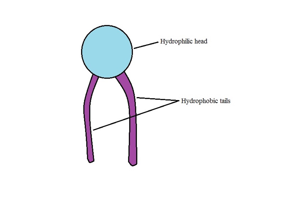
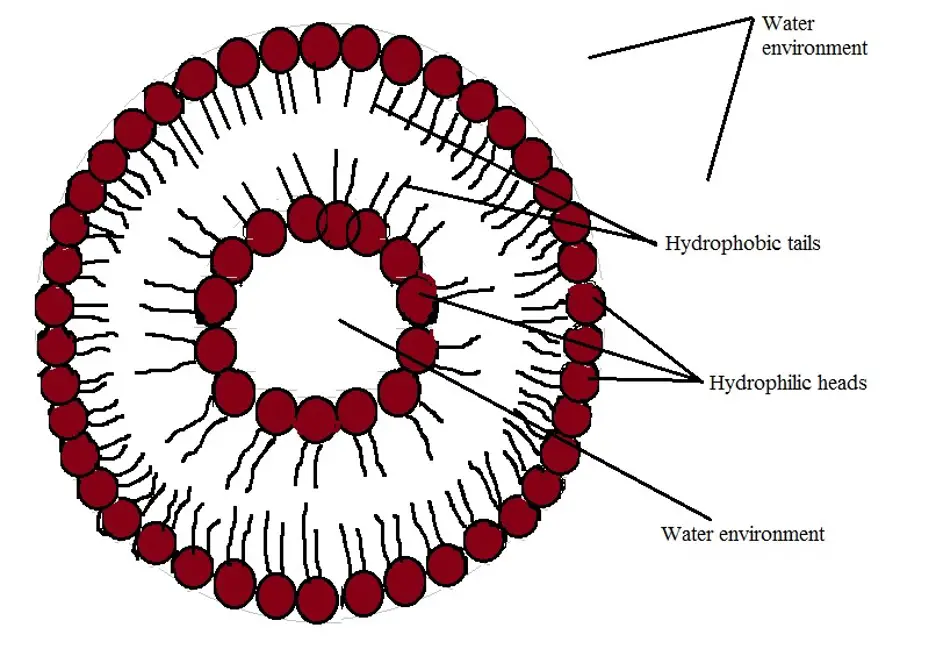
A cell membrane consists of a large number of phospholipids. As mentioned, these phospholipids consist of two distinct parts including the hydrophilic head, the part that contains the phosphate and glycerol, and the hydrophobic tail consisting of the fatty acids.
In an environment with water, the hydrophilic part interacts with water while the hydrophobic tails cluster together as they avoid interacting with water.
As a result, both the inner and outer surfaces of the membrane will consist of the hydrophilic heads that readily interact with water while the inner/central part of the membrane will consist of the hydrophobic tails that avoid interacting with water. Therefore, the cell membrane will generally appear as the diagram below
* Because of the environment in which a cell resides (consisting of water) and the nature of the cell membrane (with hydrophobic and hydrophilic parts) a hollow sphere represents the most stable structure of the membrane.
* The hydrophobic part (C-tail) consists of saturated (straight tails) and unsaturated (with a bend/kink). Whereas the saturated phospholipids tend to pack tightly, unsaturated phospholipids allow the structure to be more fluid-like by introducing spaces.
Cytoskeleton and Structural Support
While the very nature of phospholipids causes them to pack tightly in a membrane, structural support is required in order to maintain the shape of the cell as also prevents the bilayer from being damaged.
This support is provided by the cytoskeleton which is located beneath the membrane. It consists of actin filaments that form a mesh-like framework on which the membrane lies.
While the cytoskeleton is located beneath the membrane, it has been shown to interact with some of the integral membrane proteins. This not only limits the diffusion of these proteins but also enhances structural support to the membrane.
Fluid Mosaic Model of the Cell Membrane
Proposed in 1972 by S. Jonathan Singer and Garth Nicolson from the University of California, the fluid mosaic model of the cell membrane is the model used to describe the basic structure and behavior of the membrane (of many cells).
With this model, the cell membrane consists of a number of components/molecules, together make up a mosaic, that move freely in a fluid fashion. Movement of these components/molecules (proteins, cholesterol, etc) move in a manner resembling that of icebergs in the ocean.
According to studies, a number of factors influence fluidity of the cell membrane.
These include:
Temperature - As is the case with other molecules in nature (e.g. gases and liquids) an increase in temperature causes the phospholipids to grow apart and become more flexible. In the case of low temperature, the phospholipids come closer together causing the structure to become more rigid
Cholesterol - In the cell membrane, cholesterol molecules are appropriately distributed in a manner that holds phospholipids together. An increase or decrease of these molecules affects the structural integrity of the membrane as well as the ability of the membrane to control the movement of molecules and ions in and out of the cell.
Fatty acids - As already mentioned, the cell membrane consists of both saturated and unsaturated fatty acids. Because of the single bonds between the carbon atoms of saturated fatty acids, they have been shown to be straight chains that tend to pack tightly.
As a result, a membrane that only consists of these fatty acids would be solid at normal body temperature which would in turn interfere with the movement of molecules and ions in and out of the cells.
However, the presence of unsaturated fatty acids (with a double membrane between some of the carbon atoms and thus a bend/kink) creates spaces in the membrane thereby preventing the structure from being tightly packed.
By creating spaces in the membrane, unsaturated fatty acids increase fluidity of the structure and also allow some of the molecules to pass without much difficulty.
Functions
The cell membrane is a continuous sheet that separates the inner part of the cell from the external environment. As such, it serves a number of important roles that allow the cell to function normally.
These include:
Morphological/Compartmentalization - The cell membrane confines various subcellular organelles thereby separating them from the external environment. Being a continuous sheet, the membrane allows normal intracellular processes to continue without interruptions from the external environment.
Without this membrane, for instance, communication required for given molecules to be synthesized is affected thereby changing various cellular processes. In addition, the relationship between neighboring organelles (e.g. between the nucleus and ribosome) is altered.
Control the movement of material/substances in and out of the cell - Being the barrier between the cell and the external environment, the cell membrane is involved in regulating the movement/transport of various molecules and ions in and out of the cell.
Here, the transport/movement of these substances may occur through a number of mechanisms that include:
Diffusion
Essentially, diffusion involves the movement of substances from an area with high concentration to an area of low concentration. As such, this mode of movement is largely dependent on an imbalance in concentration inside and outside the cell. With regards to the movement of substances, diffusion may take three forms.
These include:
Passive or simple diffusion - As the name suggests, this type of diffusion is simple and involves the movement of molecules from where they are highly concentrated to the area of low concentration.
For the most part, these molecules are small and nonpolar. A good example of this is the diffusion of gases (e.g. oxygen) in and out of the cell.
Some of the other molecules that pass through the cell membrane are water molecules, which have a little charge, and carbon dioxide.
Facilitated diffusion - Unlike simple diffusion, facilitated diffusion is a process in which molecules have to be assisted to pass through the membrane. While it is also dependent on a concentration gradient, this mode of transport is also dependent on such channels as the protein channels, (e.g. aquaporins that transport water molecules).
Molecules that depend on this mode of transport tend to be larger and have polarity (e.g. sodium and potassium articles). Also, facilitated diffusion may occur through carrier proteins.
Here, the molecules attach to the carrier proteins which cause it to change shape in a manner that allows the molecules to be transported in or out of the cell.
In this mode of transport, the carrier proteins may only bind to a given type of molecule (specific) - Ion channels may also be included in this category. These channels are often gated and thus only allow a given type of ion.
Active transport
Unlike diffusion, active transport is the type of transport where molecules have to be transported against their gradient. As such, it's not dependent on the concentration gradient.
Given that active transport involves moving molecules against a concentration gradient, it also requires energy to do so.
There are two main types of active transport that include:
Primary active transport - A good example of primary active transport is the sodium/potassium pump (Na+/K+ pump). Here, the transporter (protein) is embedded into the membrane and extends the entire length of the membrane.
Typically, the extracellular side of this transporter is closed while the intracellular side is open. The intracellular side is associated with ATP - a source of energy. In this state, the transporter has a high affinity for sodium ions located in the cell.
These ions enter and attach to the transporter which in turn causes the ATP to transfer phosphate to the transporter. This provides the transporter with the energy that is required to close the open intracellular side and open the extracellular end.
As a result, an affinity for sodium ions reduces allowing them to be released to the extracellular environment. Once these ions are released, the transporter increases its affinity for potassium which causes potassium ions to enter and attach to the transporter. This is followed by the closing of the extracellular end while the phosphate at the intracellular end is released.
Therefore, a new ATP binds to this site causing the intracellular end to open and release the potassium into the cell. This mode of transport uses a significant amount of energy. For this reason, these transporters are few.
Secondary active transport - While secondary active transport also relies on membrane proteins, the energy used here is derived from ions electrochemical gradient rather than APT. One of the proteins involved in this mode of transportation is the sodium-glucose co-transporter 1.
Initially, the side of this protein that is in contact with the cytoplasm is closed while the extracellular end is open. In this state, two negatively charged sodium ion binding sites are exposed and attract two sodium ions (sodium anions).
As the ions move down the electrochemical gradient, energy is released, causing the protein transporter to change shape. In turn, this increases its infinity for glucose molecules located outside the cell. Glucose in this environment is in low concentration.
An increase in affinity for glucose causes glucose molecules to attach to the transporter resulting in the closing of the extracellular end of the protein as the cytoplasm end opens. With the release of sodium ions into the cell, affinity for glucose by the protein decreases allowing the glucose to be released into the cell.
Vesicle transport - The third type of transport is known as vesicle transport and involves the formation of membrane-bound vesicles. This involves such processes as endocytosis and pinocytosis where a molecule is internalized having come in contact with the cell membrane.
During endocytosis (receptor-mediated endocytosis), for instance, the substance (e.g. low-density lipoproteins) first comes in contact with the specific receptors located on the cell membrane. This stimulates the formation of vesicles on the membrane through inward bending of the membrane.
In the process, the molecule is enclosed in the vesicle and transported into the cell. Similar processes are important for the transport of molecules required by the cell, phagocytosis as well as feeding by such organisms as amoebae – This method is commonly used to transport larger molecules.
See page on Passive Diffusion Vs Active Transport
Cell signaling
As mentioned, the cell membrane consists of a variety of molecules. Some of these molecules are protein receptors that bind to specific types of molecules.
Good examples of these receptors are insulin receptors that bind insulin. By binding to given signal molecules in the extracellular environment, these receptors (transmembrane proteins) allow the cell to respond appropriately to the stimuli - Information from the stimuli is transmitted across the cell membrane and into the cell through such pathways as signal transduction cascades.
Animal Cells and Plant Cells
Both plants and animals are eukaryotes and thus have cells with membrane-bound organelles. Like animal cells, plant cells also contain various important organelles including the nucleus, mitochondria, the Golgi apparatus, and ribosomes among others. However, plant cells contain a number of extracellular components not found in animal cells. These include the chloroplast, a large central vacuole as well as the cell wall.
In plants, the cell membrane is located between the cell cytoplasm and the cell wall. As is the case with animal cells, the cell membrane in plants is a lipid bilayer. It's made up of phospholipids and also contains protein and carbohydrate molecules. Here, the cell membrane is involved in a number of functions including containing cell organelles, transportation of molecules in and out of the cell as well as cell communication.
The cell wall is a harder layer that surrounds the cell membrane. Unlike the cell membrane, the cell wall of plant cells is primarily made up of cellulose. It also consists of pectin among several other components. By surrounding the cell membrane, the cell wall serves to provide tensile strength while also protecting the cell against various environmental stresses.
In addition, it also contributes to tissue rigidity by withstanding turgor pressure - internal cell pressure that pushes the cell membrane in plant cells against the cell wall. As such, it plays an important role in limiting water loss (and thus the loss of turgor pressure) that would otherwise cause wilting.
Cell Membrane Vs Plasma Membrane
The words cell membrane and plasma membrane are often interchanged to mean the same thing. However, plasma membrane refers to the type of membrane that surrounds cell organelles while the cell membrane is the membrane that surrounds the cell as a whole. The plasma membrane is commonly found in eukaryotic cells that contain membrane-bound organelles.
In these cells, this membrane forms the barrier between the cell organelles (e.g. nucleus and mitochondria) and the external environment (external to the organelles). Like the cell membrane, however, the plasma membrane is also made up of phospholipids and also consists of proteins and carbohydrates.
In addition to being a barrier that separates the organelles from the cellular environment, the membrane is also involved in communication between cells and cell components as well as regulating the movement of molecules in and out of the organelles.
* Some of the organelles contain a double membrane (e.g. mitochondria and nucleus) while others only have a single membrane (e.g. vacuole, Golgi apparatus, and lysosome etc).
See also: What are the Functions of Lipids, Proteins and Lipopolysaccharides on the Cell Membrane?
Return to Cell Biology main page
Return from Cell Membrane to MicroscopeMaster home
References
David E. Sadava. (1993). Cell Biology: Organelle Structure and Function.
Jeremy M Berg, John L Tymoczko, and Lubert Stryer. (2002). Biochemistry, 5th edition.
Natalie Gugala, Stephana Cherak, and Raymond J. Turner. (2016). The Membrane.
Valerica Raicu and Aurel Popescu. (2008) Cell Membrane: Structure and Physical Properties. In: Integrated Molecular and Cellular Biophysics.
Links
https://www.sciencedirect.com/topics/medicine-and-dentistry/cell-membrane
https://opentextbc.ca/biology2eopenstax/chapter/lipids/
Find out how to advertise on MicroscopeMaster!
