Types of Cell Division
Definition, Mitosis, Meiosis & Vs Cancer
Introduction
Cells are the most basic units of life, and every living organism is made up of one or more cells. These cells reproduce by copying their genetic information and undergoing cell division, where the parent cell gives rise to two daughter cells.
There are three major types of cell division, which are:
- Binary fission
- Mitosis
- Meiosis
Whereas binary fission takes place in prokaryotic cells of simple single-celled organisms such as bacteria. Mitosis and meiosis take place in eukaryotic cells and are more advanced.
Although there are differences between prokaryotes and eukaryotes, there are a number of features that are common during their processes of cell division.
These include:
- The replication of DNA,
- The segregation of the original and the replica,
- Cytokinesis
Binary Fission
Binary fission is the type of cell division that takes place in prokaryotes (bacteria and archaea). Genetic material (nucleoid) in these cells is arranged in a single circular chromosome of the DNA.
In a similar fashion to eukaryotes, the genetic material of these cells is duplicated before division.
As the prokaryote elongates, the two chromosomes attached to the plasma membrane also move apart, and once the two copies have separated (the original and replicate chromosomes), the cell divides, a process referred to as cytokinesis.
Except in a case of mutation, the two resultant cells are identical.
Eukaryotes
Eukaryotic cells are more complicated given that they contain more organelles as compared to the prokaryotic cells.
The cell cycle, which is composed of four main phases, is responsible for planning and the development of these cells.
This cycle is also composed of all the steps required for the reproduction of a eukaryotic cell.
The four phases of a cell cycle include:
- G1- during this phase, a cell prepares for the synthesis stage by making sure that all material required for DNA synthesis are present.
- S phase- during the phase, DNA is copied so that the daughter cells have a complete set of chromosomes at the end of the cycle.
- G2 phase- this is the second phase of growth, and the cell is preparing for mitosis/ meiosis by making sure that all the raw material required for the physical separation are present.
- M phase- it is during this phase (mitosis) that the cell separates into two new cells.
The first three stages make up the interphase stage, where the cell spends about 90 percent of the cycle.
Mitosis
This is the process through which identical daughter cells are formed through replication and the original replication of chromosomes.
The daughter cells are identical to the parent cells in that, if the parent cell is diploid, then the daughter cell will be diploid as well.
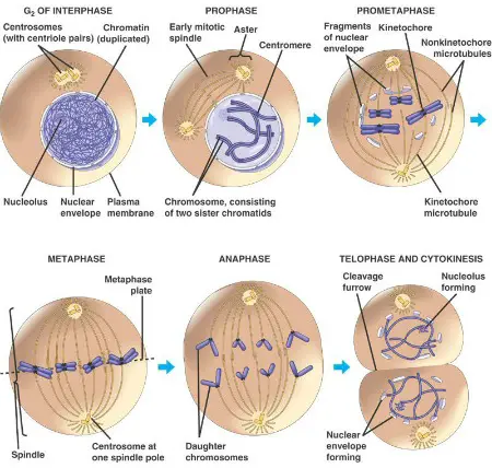
During mitosis, the replicated chromosomes are positioned at the middle of the cytoplasm. They are segregated so that the daughter cells can get a copy of the original DNA.
This is made possible by the presence of microtubules (spindle fibers), which pull the chromosomes into each of the cells.
These fibers arise from the centrioles, which are on either side of the cells, and may have even smaller microtubules referred to as aster. These are thought to serve as braces for the functioning of the fibers.
Interphase
During the interphase period, the cell replicates its DNA (chromosomes) as it prepares for the division. Chromosomes at this stage are not easily visible since they are uncoiled.
Prophase
This is the first phase in mitosis. The nuclear envelope also starts to dissolve. The chromosomes also starts coiling and spindle fibers start forming as centrosomes divide and start migrating to either side of the cell.
During the pro-metaphase the nuclear envelope breaks down and the kinetochore microtubules appear to interact with polar microtubules of the spindle fibers.
This brings about the movement of the chromosomes.
Metaphase
At this stage, chromosomes, which consist of chromatids held together by the centromere start migrating to the equator of the spindle fibers. The chromosomes therefore become aligned on a plane as the kinetochore fibers attach to the spindle fibers.
Anaphase
This phase starts with separation of centromeres thus separating the chromatids hence doubling the number of chromosomes. The new chromosomes then begin moving towards the poles of the cells
Telophase
During telophase, chromosomes reach poles of their respective spindles. The nuclear membrane also starts to reappear and chromosomes uncoil.
The spindle fibers also start breaking down and the cell divides in to two. The cells then start developing in to different adults. This phase passes the two cells in to the interphase stage.
During G1 interphase, the chromosomes have one chromatid. The chromosomes then replicates and each of them gets two sister chromatids.
Meiosis
Meiosis is involved in the production of sperm and eggs in individuals who can sexually reproduce. This process reduces chromosomes by half.
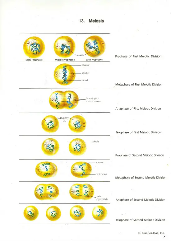
In this case, when a sperm fertilizes and egg, the zygote that results has a full set of chromosomes. A cell that undergoes meiosis therefore divides two times (meiosis 1 and meiosis 2). The diploid (2n) parent cell results in 4 haploid (n) gametes.
Meiosis 1 is known as the reduction phase while meiosis 2 is the division phase. In meiosis, unlike in mitosis, two chromosomes in a homologous pair will line up next to each other (synapsis). The resulting homologous pair is referred to as bivalent.
Interphase
This is similar to that of mitosis except for the fact that in meiosis, it is followed by two cell divisions.
Prophase 1
Chromosomes condense and attach to the nuclear envelope. Synapsis then takes place and tetrads are formed, each composed of four chromatids. Crossing over of genetic material may occur during the synapsis as the chromosomes thicken and detach from the nuclear envelope.
Like in mitosis, centrioles also move and migrate to the poles as the nuclear and nucleoli start breaking down.
Metaphase 1
In this phase, tetrads align at the metaphase plate and the centromeres of the homologous chromosomes become oriented towards either side of the cell poles.
Anaphase 1
The chromosomes start moving to the opposite poles of the cells. The microtubules and kinetochore fibers also interact and there is movement. Unlike in mitosis, homologous chromosomes move to the opposite poles but sister chromatids remain together.
Telophase 1
The spindle fibers continue moving homologous chromosomes to the poles after which either pole has a haploid number of chromosomes. Cytokines also tend to occur spontaneously.
Two daughter cells are formed each containing a half number of chromosomes as the original parent cell.
Meiosis 2
Prophase 2
In this stage, the nuclear membrane as well as the nuclei break and the spindle fibers reappear. The chromosomes then start migrating to the equator of the cell.
Metaphase 2
Chromosomes line up at the second metaphase plate at the center of the cell. The kinetochores then point towards the opposite poles of the cell.
Anaphase 2
The sister chromatids divide and move to the opposite poles of the cells.
Telophase 2
In this phase, distinct nuclei start forming at the opposite pole and cytokines start occurring. At the end of this phase, there are four daughter cells. Each of these has half the number of the chromosomes in the original parent cell.
Cancer Cell Division
Typically, cancer is a disease of mitosis. In this case, the normal checkpoints that regulate mitosis are overridden by the cancerous cells.
This starts in an event where a single cell transforms from a normal cell in to a cancerous one as a result of the changes in functions on one of a number of genes that play a role in controlling growth.
Cancer cells tend to ignore the normal growth density dependent inhibition. They then multiply following contact with other cells and continue piling up until all the nutrients are exhausted.
Cancer: Unregulated Cell Division - Animation
See also articles about cytopathology and histopathology
Conclusion
In conclusion, it is important to keep in mind that there are three types of cell division, each of which plays a special purpose and achieves a given goal.
Mitosis, being the most common form of cell division is important for growth and repair since cells wear out and have to be replaced. Mitosis is also important for the repair of damaged tissues and allows for growth.
Meiosis, which is a unique form of cell division in eukaryotes, is important for the formation of gametes, which are required for reproduction and continuation of species. For simpler organism such as bacteria, binary fission, which is a form of asexual reproduction allows for the continuation of the organisms.
However, this is not the only type of asexual reproduction. In understanding cell division and mitosis in particular, researchers have come to understand cancers, which has led to new and targeted treatments that are constantly being enhanced.
Check out information regarding prokaryotes and eukaryotes
Also, an interesting read on protists
Molecular Biology of the Cell 5th Edition reviewed and an advanced article on Cytochemistry.
Learn more about cells in these articles: Cell Culture, Cell Differentiation, Cell Staining, Tissue Culture, Time-Lapse Microscopy, the different organelles as well as cell apoptosis.
Read about Onion Root Tip Mitosis
Why is Cell Adhesion important?
What are the differences between Meiosis and Mitosis?
Take a closer look at the Nucleus
Return to Cell Biology Main Page
Return to learning about Binary Fission
Return from Cell Division to Microscopy Research at MicroscopeMaster Home
Find out how to advertise on MicroscopeMaster!


