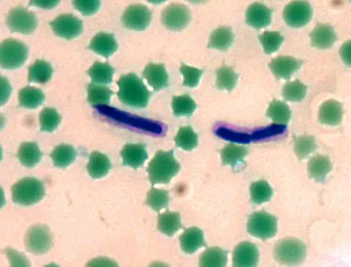
Capsule Stain
Definitions, Methods and Procedures
What is a Capsule?
Some bacteria like Bacillus anthracis have a well organized layer of material lying outside the cell wall referred to as a capsule (or K antigen). What really differentiates a capsule from any other material that may form on the surface of a cell wall of other bacteria is the fact that capsules are well organized layers that cannot be easily washed off.
Typically, a capsule is composed of capsular polysaccharides (CPS) which are polysaccharides. They may also be composed of a number of other materials such as poly-D-glutamic acid. For some bacteria, capsules are very important in that they are a major virulence factor.
As such, they protect the bacteria from phagocytic actions of such cells as neutrophils allowing the bacteria to thrive. This is achieved due to the fact that the capsule is very smooth and has a negative charge that prevents attachments/adherence.
Some of the other significance of a capsule include:
- Protecting bacteria from desiccation
- Providing a food reserve
- Protecting anaerobes from oxygen toxicity
- To help bacteria attach onto surfaces
Some of the organisms that are likely to have a capsule in place include:
- Streptococcus pneumoniae,
- Haemophilus influenzae,
- Neisseria meningitidis,
- Klebsiella pneumoniae,
- Escherichia coli,
- Group B streptococci
Capsule Stain
Capsule stain is a type of differential stain which involves the use of two stains; primary stain and the counterstain. Here, it's worth noting that for most part, capsules are non-ionic.
As a result, acidic and basic stains will often fail to adhere on to them. For this reason, it becomes necessary to stain the background using an acidic stain while the cell is stained using a basic stain.
Two of the most commonly used methods used in capsule staining include:
- India ink method
- Anthony's stain method
India Ink Method
Requirements
- India ink
- Crystal violet
- Glass slide
- Inoculating loop
Procedure
- Place a drop of India ink onto a clean glass slide
- Using a sterile loop, obtain a sample and place it onto the slide and mix it with the drop of India ink
- Obtain another sterile glass slide and having laid it at an angle on one end of the first slide, spread out the drop into a film to have a thin layer of the smear-ink mixture
- Allow the slide to stand for about 5 minutes (air- dry)
- Saturate the slide with crystal violet for a minute and then tilt the slide to drain excess dye
- Allow the slide to dry (air-dry)
- View the slide using 100x
India ink is often used as the negative stain.
However, some of the other stains that can also be used include:
- Congo red
- Nigrosin
- Eosin
This is a negative staining technique that is essentially used to identify the presence of capsules. Because of its acidic nature, India ink (or Congo red, nigrosin) stains the background dark.
On the other hand, crystal violet is used for number of reasons including:
- To act as a fixative
- Increase penetration power
- Stain the cells (being a basic dye)
- Decrease pH of smear
When viewed under the microscope, the background will be dark as a result of India ink, bacteria cells will be purple having taken the crystal violet dye while the capsule will be clear against a dark background given that it takes no stain.
* Nigrosin is recommended to India ink because it gives a more even background and spreads more easily.
Anthony's Stain Method
With Anthony’s stain method, the primary stain used is crystal violet.
Some of the other requirements include:
- A sample (36-48 hour culture of capsulated bacteria)
- Glass slide
- Inoculating loop
Procedure
- Introduce a drop of crystal violet on to a clean glass slide
- Using a sterile loop, place the sample on to the glass slide
- Obtain another glass slide and at an angle, spread the drop and sample to form a thin film
- Allow the film to dry (air dry) for about 6 minutes
- Title the slide and rinse with 20% copper sulfate solution
- Allow the side to air dry for about 3 minutes
- Place the slide on to the microscope stage and observe using oil immersion
This is a positive staining method used in capsule staining. Here, the smear/sample is treated with a hypertonic solution (copper sulfate) for the purposes of creating an ionic difference that ultimately results in the diffusion of a stain towards the outer surface of the cell.
After the slide is air dried, it becomes possible to observe the stain that remained on the capsular layer. Here, one will see a dark violet color of the cell and a light violet color of the capsule.
Notes
Unlike endospores, capsules are more delicate. For this reason, using heat during the staining process should be avoided given that capsules would be easily destroyed. Rinsing the slide with water is also avoided in capsule staining because it would dislodge the capsule.
A drop of serum may be used on the smear to enhance the size of the capsule. This helps make it more visible to the technician under the typical light microscope.
Given that this is a sensitive procedure, sterile technique should be used when obtaining the sample and introducing it onto the slide. This prevents any contamination and helps determine whether the sample really has any bacteria capsules.
Significance of Capsule Stain
As previously mentioned, capsules are considered to be the virulence factor which means that they enhance the ability of a bacterial to cause a disease. With a capsule in place, the bacteria is protected from such body defense cells as macrophages. Therefore, it becomes easier for the bacteria to thrive and cause diseases. For this reason, capsule stain is of great benefit in that it helps identify the presence of capsules and thus allowing healthcare professionals to recommend the right treatment.
Related and Interesting articles:
Gram Staining - Purpose, Procedure and Preparation
Endospore Stain - Understanding Definitions, Techniques and Procedures
Learn more about Cell Culture, Cell Division, Cell Differentiation and Cell Staining.
Return from Capsule Stain to MicroscopeMaster Research Home
References
Roxana B. Hughes and Ann C. Smith . Capsule Stain Protocols. Publication Date : September 2007. http://www.austincc.edu/microbugz/capsule_stain.php
http://web2.uwindsor.ca/courses/biology/fackrell/Methods/Capsule_Stain.htm
Find out how to advertise on MicroscopeMaster!


