Basophils in Microscopy
Procedure, Staining and Observations
Basophils comprise less than 1 percent (about 0.5 percent) of the total leukocytes in circulation. They, therefore, make up the smallest percentage of white blood cells when compared to the others. Like mast cells, basophils are derived from CD34+ located in the bone marrow and have granules that contain histamine. Once fully developed and mature in the marrow, basophils then enter the circulation (blood circulation).
Although they make up the smallest percentage of leukocytes, they carry the highest amount of histamine, which is particularly important in immune response to foreign particles. They also contain heparin, which is an important anticoagulant that keeps blood from clotting too fast.
Microscopy
Basophils can be viewed by simply preparing a thin film and viewed under a phase contrast microscope. Here, students may also prepare a thick film slide for comparison.
Thin Film Preparation
Requirements
- A prick needle
- A clean glass slide (2 or 3)
- Stain (toluidine blue, methyl violet or methylene blue) - either stain can be used
- Cotton swab and alcohol
Procedure
- Using a cotton swab with alcohol, clean the tip of the middle or ring finger and prick with a clean pricking needle
- Press the finger tip and wipe off the first drop of blood
- Press the finger tip again to place a drop or two of blood on to a microscope glass slide
- Using another clean glass slide or cover slip at an angle, spread the blood drop on the glass slide to create a thin film of blood
- Allow the slide to dry off and fix with methanol before staining
- Allow the slide to dry completely
Staining
- Once the slide is dry, stain it by immersing in a jar of the stain (toluine blue, methyl violet or methylene blue) for between 20 and 30 minutes
- View under the microscope starting with low power
- At high power (1000x) add oil immersion to view
Observation
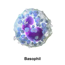
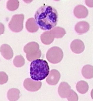
When viewed under the microscope, basophils may appear spherical in shape. However, with either of the stains mentioned, the granules stain brick red and appear as so. They are also highly refractive and when viewed under the microscope, they may seem like vacuoles because of the inverted phase contrast and the fact that they are ringed by a dark boarder.
Discussion - Basophils
When viewed under the phase contrast and electron microscope, basophils have been shown to contain granules (measuring about 0.2 um in diameter). This therefore makes them granulocytes putting them in the same category with neutrophils and eosinophils.
They have also been shown to have a diameter ranging from 10 um to 14 um making them the smallest in the category (granulocytes). However, unlike neutrophils and eosinophils, basophils are not phagocytes. There numerous, opaque granules also make it difficult to clearly see the nucleus. Unstained, it is possible to get a better view of the nucleus which has two lobes.
Some of the granules are secretory in nature and store histamine and heparin (as well as a number of other chemicals). During allergic reactions as well as in the event of inflammations, histamine is released causing increased permeability of the capillaries.
Histamine contributes to the inflammatory response by causing swelling (edema). Inflammation also results in the release of heparin, which not only prevents blood clotting as mentioned, but also activates an enzyme known as lipoprotein lipase which serves to degrade triglycerides in blood.
Histamine is also an important vasodilator, which allows for increased blood flow to tissues. This is particularly important during infections given that it allows for blood, containing other leukocytes and material to reach the affected tissue.
See Also: Lymphocytes, Neutrophils, Monocytes, Eosinophils, Mast Cells
Return to White Blood Cells Main Page
Return to Cell Biology
Return from Basophils to MicroscopeMaster Home
Find out how to advertise on MicroscopeMaster!


